
This chapter covers the Cell Cycle from the 2nd Year (FSc Part-II) Biology syllabus of the Punjab Curriculum and Textbook Board (PTB/PCTB). A cell undergoes a regular sequence of changes involving a period of growth, replication of DNA, and cell division, together called the cell cycle, comprising interphase (the period of non-apparent division) and the mitotic phase (the period of division); each of these phases is further subdivided into distinct sub-phases. These notes are prepared by freebooks.pk.
The chapter covers interphase (G1, S and G2 phases), mitosis (karyokinesis with its prophase, metaphase, anaphase and telophase stages, followed by cytokinesis, including differences between plant and animal cells), the importance of mitosis and the biology of cancer, meiosis (the substages of prophase I, and meiosis I and II, plus its importance), meiotic errors (non-disjunction and the resulting syndromes such as Down's, Klinefelter's and Turner's), and finally the two forms of cell death, necrosis and apoptosis.
Learning Objectives
- Define the cell cycle and describe the sub-phases of interphase (G1, S, G2, G0).
- Describe the stages of mitosis (prophase, metaphase, anaphase, telophase) and cytokinesis, including plant-animal differences.
- Explain the importance of mitosis and describe how uncontrolled cell division leads to cancer.
- Describe the substages of prophase I of meiosis (leptotene through diakinesis) and the role of crossing over.
- Describe meiosis I and meiosis II and explain the biological importance of meiosis.
- Define chromosomal non-disjunction and describe Down's, Klinefelter's and Turner's syndromes.
- Differentiate necrosis and apoptosis as forms of cell death.
Key Concepts
The Cell Cycle and Interphase
A cell undergoes a regular sequence of changes involving a period of growth, DNA replication, and cell division, together called the cell cycle, comprising two main phases: interphase, the period of non-apparent division, and the mitotic phase, the period of actual division. Interphase, sometimes misleadingly called the 'resting phase', is actually a period of intense biochemical activity, divided into three sub-phases. G1 (Gap 1) is a period of extensive metabolic activity in which the cell grows in size, synthesises specific enzymes, and accumulates DNA base units for upcoming DNA synthesis; a post-mitotic cell can exit the cycle during G1 into a resting state called G0, remaining there for days, weeks, or (in cells such as nerve cells and eye lens cells) even the organism's entire lifetime without further division. G1 is followed by the S-phase (synthesis phase), during which DNA is replicated (chromosomes duplicated), which then initiates G2 (the pre-mitotic phase), during which the cell prepares further for division by storing energy for chromosome movement and synthesising mitosis-specific proteins, RNA, and microtubule subunits for the spindle fibres.
At each stage of the cell cycle, specific checkpoints determine whether the cell proceeds to the next phase based on its internal state, and the length of each phase varies considerably between cell types: in a typical human cell, the full cycle takes about 24 hours, with mitosis taking about 30 minutes, G1 about 9 hours, S-phase about 10 hours, and G2 about 4.5 hours, while the full cycle in yeast cells takes only about 90 minutes.
Mitosis: Karyokinesis (Prophase through Telophase)
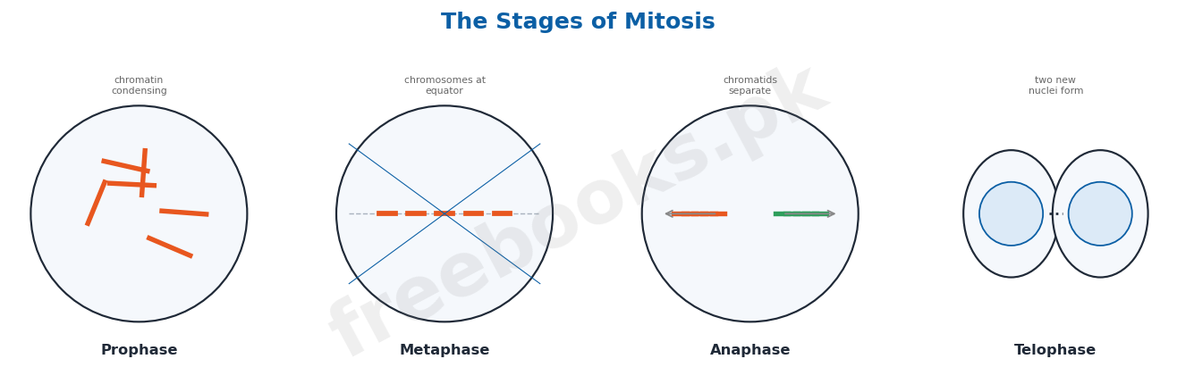
Mitosis is the type of cell division that ensures the daughter cells receive the same number of chromosomes as the parent cell; it occurs in haploid or diploid cells throughout nearly all parts of the body as needed, and although a continuous process, it is conventionally divided into karyokinesis (division of the nucleus) and cytokinesis (division of the whole cell). At the start of mitosis in an animal cell, the centriole pairs (already duplicated during interphase) separate and migrate to opposite sides of the nucleus, establishing bipolarity; three sets of microtubules radiate from each pair of centrioles – astral microtubules (forming the aster), kinetochore microtubules (attaching to chromosomes at their kinetochores), and polar microtubules (interdigitating with polar microtubules from the opposite pole) – together forming the mitotic apparatus, a structure larger than the nucleus, designed to capture, align and separate chromosomes to ensure their equal distribution. Karyokinesis is itself conventionally divided into four stages. During prophase, the fine chromatin network condenses into visible, increasingly thick chromosomes (each showing two sister chromatids joined at the centromere), the nuclear envelope and nucleoli disappear, releasing nuclear material into the cytoplasm, and the mitotic apparatus organises as the cytoplasm becomes more viscous.
During metaphase, each duplicated chromosome (two sister chromatids joined at the centromere, which bears a specialised kinetochore region) has kinetochore fibres from each spindle pole attach to its kinetochore, aligning all the chromosomes at the equator of the spindle to form the metaphase (equatorial) plate. Anaphase is the most critical phase, ensuring equal distribution of chromatids: kinetochore fibres contract toward their poles while polar microtubules elongate, separating the sister chromatids at the centromere so that half travel to each pole. Telophase begins once the chromosomes reach the poles: they decondense back into chromatin, the mitotic apparatus disassembles, and the nuclear membrane and nucleoli reorganise around each set of chromosomes, forming two new nuclei. During late telophase, cytokinesis begins as astral microtubules signal the cell's equatorial region, activating actin and myosin to form a contractile ring, which produces a cleavage furrow that deepens inward, ultimately dividing the parent cell into two daughter cells.
Mitosis in Plant Cells and the Importance of Mitosis
Mitotic events in plant cells broadly resemble those in animal cells but show important differences: most higher plants lack visible centrioles, instead having an analogous region from which spindle microtubules radiate, and the rigid cell wall prevents the plant cell from changing shape as dramatically as an animal cell does during division. At cytokinesis, instead of a contractile ring, plant cells form a membrane structure called the phragmoplast, built from Golgi-derived vesicles that originate during metaphase, line up at the cell's centre, and fuse at the end of telophase; these vesicle membranes become the new plasma membrane of the daughter cells, while their contents (including cellulose and pectin precursors) form the new cell wall material.
Mitosis is essential because it equally distributes hereditary material to daughter cells; since there is no crossing over or recombination, genetic information remains unchanged from generation to generation of cells, ensuring continuity. Mitosis underlies asexual reproduction in many plants and animals, regeneration, wound healing, replacement of aging cells, and the orderly growth and development of multicellular organisms, and it is also essential for tissue culture and cloning techniques; because of its importance, mitosis must be carefully managed and controlled, since malfunction can result in tumours and lethal diseases such as cancer.
Cancer: Uncontrolled Cell Division
Normal cell multiplication is carefully regulated and responsive to the body's specific needs, balancing cell death and birth to maintain a steady state; when this regulatory control breaks down, a cell may begin growing and dividing in an unregulated fashion without regard for the body's actual need for more cells of its type, and its unrestrained proliferation produces an unwanted clone of cells called a tumour, which can expand indefinitely. Tumours are of two basic types: benign tumours are small, localised, and generally behave like normal cells with little harmful effect (except through physical interference or hormone-like secretions), while malignant tumours (cancers) divide more rapidly, invade surrounding tissue, enter the circulatory system, and establish secondary growth sites away from the original tumour, a process called metastasis. Cancer cells can be distinguished from normal cells by being less differentiated, showing characteristics of rapid growth such as a high nucleus-to-cytoplasm ratio, prominent nucleoli, and frequent mitosis, with invading cells in otherwise normal tissue indicating malignancy.
Cancer is caused mainly by mutations in somatic cells, typically accumulating from as few as three to as many as twenty mutations in genes that regulate cell division; these mutations produce two fundamental changes in cancer cells: first, metastatic cells break contact with neighbouring cells and overcome the normal restrictions on cell movement imposed by the basal lamina and other barriers, allowing them to invade other body regions; second, they proliferate without limit, ignoring the body's normal regulatory checks and programmes.
Meiosis I: Prophase I and the Significance of Crossing Over
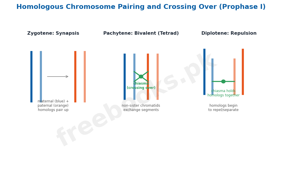
Meiosis is a specialised type of cell division that reduces the chromosome number in daughter cells to half that of the parent cell, occurring during gamete formation in animals and spore formation in plants; because it involves two consecutive divisions (meiosis I and meiosis II) after a single round of DNA replication, each diploid cell produces four haploid cells. Meiosis I is the reduction division, while meiosis II resembles mitosis; both are divided into prophase, metaphase, anaphase and telophase stages (numbered I and II respectively). Prophase I is a notably prolonged phase that differs from mitotic prophase because homologous chromosomes (similar but not necessarily identical chromosome pairs, one from each parent) pair up; it is itself divided into five substages. In leptotene, chromosomes become visible, shorten and thicken, and homologous chromosomes begin approaching each other. In zygotene, synapsis (the highly specific pairing of homologous chromosomes) begins, forming a paired structure called a bivalent or tetrad. In pachytene, pairing is completed, chromosomes thicken further, and each bivalent (with four chromatids) undergoes crossing over, in which non-sister chromatids exchange segments at points called chiasmata, reshuffling genetic material and producing recombination; pachytene may last days, weeks, or even years, while leptotene and zygotene last only a few hours.
In diplotene, the paired homologous chromosomes begin to repel and separate from one another, though separation remains incomplete since they stay joined at their chiasmata (points of exchange). In diakinesis, chromosome condensation reaches its maximum, the separation begun in diplotene completes (though the chromosomes typically remain joined at one point, often near the ends), and the nucleoli disappear. In metaphase I, the nuclear membrane disassembles, spindle fibres form, and kinetochore fibres attach to the kinetochore of each homologous chromosome from opposite poles, aligning the bivalents at the equator, with the two sister chromatids of each chromosome behaving as a single unit. In anaphase I, unlike mitotic anaphase, sister chromatids remain joined: kinetochore fibres contract and polar fibres elongate, pulling each whole chromosome (still with two chromatids) to its respective pole, so each pole receives half the total chromosome number – the actual reduction step. In telophase I, the nuclear membrane and nucleoli reform around each set of chromosomes, producing two nuclei each with half the original chromosome number, after which the cytoplasm divides, completing the first meiotic division; chromosomes may decondense at this stage.
Meiosis II and the Importance of Meiosis
After telophase I, the two daughter cells undergo a brief interphase, but unlike mitotic interphase, no further chromosome replication occurs. Prophase II, metaphase II, anaphase II and telophase II closely resemble the corresponding mitotic phases: chromosomes condense, the mitotic apparatus forms, chromosomes align at the equator, and sister chromatids separate and move to opposite poles, ultimately producing four nuclei (two from each of the two cells formed after meiosis I); cytokinesis then produces four haploid cells, each with half the original chromosome number.
Meiosis's importance rests on two key phenomena: crossing over, in which parental chromosomes exchange segments to produce a large number of new gene combinations (recombinations), and the random assortment of homologous chromosomes during anaphase I, which produces a very wide variety of possible gamete combinations; together these processes generate genetic variation, providing both the raw material for evolution and the basis for each individual's unique characteristics, meaning even siblings from the same parents are never genetically identical. Meiosis occurs during gamete or spore formation, halving the chromosome number so that it is restored to the normal diploid number upon fertilization, keeping the chromosome number constant across generations; without meiosis, chromosome number would double with every generation, an unsustainable outcome.
Meiotic Errors: Non-disjunction and Chromosomal Syndromes
Although meiosis is normally an orderly process, errors sometimes occur; one important abnormality is chromosomal non-disjunction, in which chromosomes fail to segregate properly during anaphase and telophase, resulting in an unequal distribution of chromosomes among daughter nuclei and causing an increase or decrease in chromosome number, with serious physical, social and mental consequences; non-disjunction may affect autosomes or sex chromosomes. Down's syndrome (mongolism) results from autosomal non-disjunction of chromosome 21, producing a gamete with 24 chromosomes that, upon fertilization with a normal gamete, yields an individual with 47 chromosomes (trisomy 21, 2n+1); it appears to occur mainly in the ovum and is strongly linked to maternal age (roughly 1 in several thousand for teenage mothers, 1 in 100 by age 40, and about three times greater risk by age 45), producing a flat, broad face, squint eyes with an inner eyelid fold, a protruding tongue, mental retardation, and defective central nervous system development; non-disjunction of chromosomes other than 21 usually causes miscarriage or very early death.
Klinefelter's syndrome results from an additional sex chromosome, typically 47 chromosomes (44 autosomes + XXY); affected individuals are phenotypically male but frequently show enlarged breasts, tendency toward tallness and obesity, small testes with no sperm production, and underdeveloped secondary sexual characteristics (rarer variants include XXXY, XXXXY and XYY karyotypes). Turner's syndrome results from a missing X chromosome, giving only 45 chromosomes (44 autosomes + X); many affected pregnancies do not survive to term, while those that do survive have a female appearance with short stature, a webbed neck, absent ovaries, and complete absence of germ cells.
Necrosis and Apoptosis: Two Forms of Cell Death
Cells rely on various extracellular and intracellular signals to regulate their activities, such as division, differentiation, morphogenesis and motility, and even a cell's death can be a predetermined, programmed event. Programmed cell death is essential for properly controlled multicellular development, sometimes eliminating an entire structure (such as the tail of a developing human embryo) or part of one (such as the tissue between developing fingers), and also regulates cell numbers, since most neurons in the human body actually die during normal development. Cell death in multicellular organisms occurs in two fundamentally different ways: a cell may commit 'suicide' in the absence of survival signals (trophic factors), or it may be actively 'murdered' by killing signals from other cells.
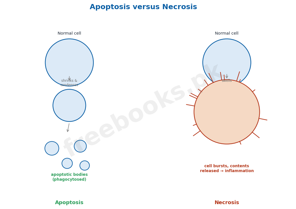
The internal programme of events and morphological changes by which a cell commits suicide is collectively called apoptosis (from the Greek for 'dropping off' or 'falling off'); during apoptosis, the dying cell shrinks and condenses, eventually splitting into small, membrane-bound apoptotic bodies that are generally engulfed (phagocytosed) by neighbouring cells, so intracellular contents are never released freely into the surrounding tissue, avoiding damage to neighbouring cells. In contrast, necrosis is cell death resulting from tissue damage, during which the dying cell swells and bursts, releasing its intracellular contents into the surrounding tissue, which can damage neighbouring cells and trigger inflammation.
Important Definitions
Cell cycle
The regular sequence of changes a cell undergoes, comprising growth, DNA replication, and cell division, divided into interphase and the mitotic phase.
Interphase
The period of the cell cycle between two consecutive divisions, comprising G1, S and G2 phases, characterized by intense biochemical activity.
Mitosis
The type of cell division that ensures daughter cells receive the same number of chromosomes as the parent cell.
Synapsis
The highly specific pairing of homologous chromosomes that occurs during zygotene of meiotic prophase I.
Crossing over
The exchange of chromosome segments between non-sister chromatids of homologous chromosomes during pachytene, producing genetic recombination.
Non-disjunction
The failure of chromosomes to segregate properly during anaphase and telophase, resulting in an abnormal chromosome number in daughter cells.
Apoptosis
The internally programmed sequence of events by which a cell commits controlled 'suicide,' shrinking and fragmenting into membrane-bound bodies that are phagocytosed.
Necrosis
Cell death resulting from tissue damage, in which the cell swells and bursts, releasing its contents and potentially damaging neighboring tissue.
Key Facts
| Item | Fact |
|---|---|
| Human cell cycle timing | Average human cell cycle ≈ 24 hours: G1 ≈ 9 hrs, S-phase ≈ 10 hrs, G2 ≈ 4.5 hrs, mitosis ≈ 30 minutes. |
| Yeast cell cycle timing | Full cell cycle in yeast cells takes only about 90 minutes. |
| Meiosis chromosome outcome | One diploid (2n) cell after meiosis produces 4 haploid (n) cells (2 divisions after 1 DNA replication). |
| Down's syndrome (trisomy 21) | Down's syndrome = 47 chromosomes (2n+1), non-disjunction of chromosome 21. |
| Klinefelter's syndrome | Klinefelter's syndrome = 47 chromosomes (44 autosomes + XXY). |
| Turner's syndrome | Turner's syndrome = 45 chromosomes (44 autosomes + X, one X missing). |
| Down's syndrome maternal age risk | Risk of Down's syndrome: ~1 in several thousand (teenage mother) -> ~1 in 100 (age 40) -> ~3x greater by age 45. |
| Cancer mutation accumulation | Cancer typically results from accumulation of about 3 to 20 mutations in cell-division-regulating genes. |
Diagrams & Illustrations
The Stages of Mitosis: a panel diagram showing the sequential stages of mitosis in an animal cell: prophase (chromosomes condense), metaphase (chromosomes align at the equatorial plate), anaphase (sister chromatids separate to poles), and telophase (two new nuclei form).
Homologous Chromosome Pairing and Crossing Over in Meiosis I: a diagram showing synapsis of homologous chromosomes to form a bivalent (tetrad) during zygotene/pachytene, and crossing over between non-sister chromatids at a chiasma during prophase I.
Apoptosis versus Necrosis: a comparative diagram showing apoptosis (cell shrinks, condenses, and fragments into phagocytosed apoptotic bodies) alongside necrosis (cell swells and bursts, releasing contents and causing inflammation).
Short Questions & Answers
Differentiate G1, S and G2 phases of interphase.
G1 is a period of metabolic activity and cell growth in which enzymes are synthesized and DNA precursors accumulate; the S-phase is when DNA is actually replicated (chromosomes duplicated); G2 is the pre-mitotic phase in which the cell synthesizes mitosis-specific proteins, RNA and microtubule subunits in preparation for division.
What is the mitotic apparatus, and what is its function?
The mitotic apparatus is the specialized microtubule structure (aster + spindle) formed from centriole-derived microtubules during mitosis; it attaches to and captures chromosomes at their kinetochores, aligns them at the metaphase plate, and separates them to ensure equal chromosome distribution to daughter cells.
How does mitosis differ between plant and animal cells?
Most higher plants lack visible centrioles (having an analogous spindle-organizing region instead), plant cell shape changes little during division due to the rigid cell wall, and plant cytokinesis forms a phragmoplast from Golgi-derived vesicles (rather than an animal cell's actin-myosin contractile ring), which becomes the new plasma membrane and cell wall material.
Differentiate anaphase I of meiosis from anaphase of mitosis.
In mitotic anaphase, sister chromatids separate and move individually to opposite poles; in anaphase I of meiosis, sister chromatids remain joined and whole homologous chromosomes (each with two chromatids) separate and move to opposite poles, which is the actual chromosome-number-reducing step.
What is non-disjunction, and give one example of a resulting syndrome.
Non-disjunction is the failure of chromosomes to segregate properly during anaphase/telophase, causing an abnormal chromosome number in daughter cells; Down's syndrome (trisomy 21, 47 chromosomes) is a well-known example, caused by non-disjunction of chromosome 21.
Differentiate apoptosis and necrosis.
Apoptosis is programmed cell 'suicide' in which the cell shrinks, condenses, and fragments into membrane-bound apoptotic bodies that are phagocytosed without releasing contents; necrosis is cell death from tissue damage in which the cell swells and bursts, releasing its contents and causing inflammation and damage to neighboring cells.
Long Questions & Answers
Describe in detail the four stages of karyokinesis (prophase, metaphase, anaphase and telophase) during mitosis.
Mitosis, though a continuous process, is conventionally divided into four distinct stages for study, and it begins with prophase, during which the fine, thread-like chromatin network that is invisible during interphase begins to condense through progressive folding, gradually becoming visible as increasingly thick chromosome structures ranging from about 0.25 to 50 micrometres in length; as prophase advances, each chromosome becomes clearly visible as a duplicated structure consisting of two sister chromatids joined together at a single point called the centromere, and toward the end of prophase several other major changes occur simultaneously – the nuclear envelope breaks down entirely and releases the nuclear material directly into the surrounding cytoplasm, the nucleoli disappear, the mitotic apparatus (comprising the aster and spindle, built from microtubules radiating out from the now-separated centriole pairs at opposite poles of the cell) becomes fully organized, and the cytoplasm itself becomes noticeably more viscous in preparation for the mechanical work of chromosome separation that is to follow. The cell then enters metaphase, in which each chromosome, still consisting of its two sister chromatids joined at the centromere, becomes attached to the mitotic spindle through its kinetochore, a specialized region located at the centromere with a particular arrangement of DNA sequences and associated proteins specifically adapted to bind the kinetochore microtubules of the spindle apparatus; critically, each kinetochore receives exactly two microtubule fibres, one originating from each of the two opposite spindle poles, and this precisely balanced, bipolar attachment is what allows all of the chromosomes in the cell to become aligned together along a single plane at the very centre of the spindle, roughly equidistant between the two poles, forming what is called the equatorial plate or metaphase plate, a configuration that must be achieved correctly before the cell is permitted to proceed further. Anaphase, widely regarded as the single most critical and consequential phase of the entire mitotic process, follows immediately once every chromosome has become properly and completely attached to spindle fibres from both poles; during this phase, the kinetochore fibres of the spindle apparatus begin to actively contract and shorten, physically pulling toward their respective poles, while at the very same time the polar microtubules, which run between the two poles rather than attaching to chromosomes, begin to elongate and push the two poles further apart, and the combined mechanical effect of these two simultaneous processes generates enough force to physically break the connection joining each pair of sister chromatids at the centromere, so that the two sister chromatids of every chromosome are pulled cleanly and completely apart from one another, with exactly one full set of chromatids, now considered individual daughter chromosomes in their own right, travelling steadily toward each of the two opposite poles of the dividing cell, thereby guaranteeing that each future daughter cell will receive a genetically complete and identical set of chromosomes. Once the separated chromosomes have fully reached their respective poles at opposite ends of the cell, anaphase is considered complete and the cell enters the final stage, telophase, during which the entire sequence of structural changes seen earlier during prophase is essentially reversed in order to reconstruct two separate, fully functional nuclei: the tightly condensed chromosomes at each pole begin to decondense and unfold, gradually losing their compact, visible chromosome structure and reverting back into the loosely dispersed, essentially invisible chromatin form characteristic of interphase, while simultaneously the mitotic apparatus itself completely disassembles and disappears, a brand new nuclear membrane reforms and reorganizes around each of the two separated sets of decondensing chromosomes, and the nucleoli, which had disappeared back during prophase, now reappear within each newly forming nucleus – together, these coordinated changes result in the formation of two genetically identical, fully reconstituted nuclei positioned at opposite poles of what remains, at this point, still a single, undivided cell, setting the stage for the subsequent physical division of the cytoplasm itself during cytokinesis, which finally separates this one cell into two completely independent daughter cells.
Describe the substages of prophase I of meiosis and explain the biological significance of crossing over and random chromosome assortment.
Prophase I of meiosis is a notably prolonged and considerably more elaborate phase than the corresponding prophase of ordinary mitosis, distinguished above all by the fact that homologous chromosomes, meaning the two versions of each type of chromosome present in a diploid cell, one originally inherited from the mother and one from the father, actively come together and pair with one another in a highly organized way, and for the purposes of detailed study this single extended phase is conventionally broken down into five sequential and clearly recognizable substages. The first of these, leptotene, is the stage during which chromosomes, previously invisible as diffuse chromatin during interphase, first become visible as they progressively shorten and thicken, while at the same time the overall size of the nucleus itself increases and the various homologous chromosome pairs present in the cell begin gradually moving closer to one another in preparation for the pairing process that is about to follow. This leads directly into zygotene, the stage in which the single most essential defining event of meiosis actually begins to take place: the highly specific pairing of each homologous chromosome with its partner, a precise process known as synapsis, which despite being remarkably exact in how closely and completely the two homologous partners eventually align is nevertheless not restricted to any single fixed starting point along the chromosome's length, and once this pairing process is underway, each resulting paired (though still physically distinct and unfused) structure formed by the two homologous chromosomes together is specifically referred to as a bivalent, or alternatively a tetrad, in recognition of the fact that it ultimately contains four individual chromatids in total. The cell then progresses into pachytene, the substage during which the pairing of homologous chromosomes that began in zygotene finally reaches full completion, while the chromosomes themselves continue to become progressively thicker and more condensed, and it is specifically during this pachytene stage that each fully formed bivalent, now clearly containing four tightly wrapped chromatids in total, undergoes the single most genetically consequential event of the entire meiotic process: individual non-sister chromatids belonging to the two different homologous chromosomes physically exchange corresponding segments of DNA with one another at specific points of contact called chiasmata, in a precisely regulated process known as crossing over, which has the critical effect of extensively reshuffling and recombining the genetic material originally inherited separately from the two parents, thereby generating substantial new genetic recombination; notably, this particular pachytene substage, unlike the comparatively brief leptotene and zygotene stages that typically last only a matter of hours, can itself persist for an extended and highly variable length of time, ranging anywhere from several days to several weeks or, in certain organisms and circumstances, even for a period of years. Following pachytene, the cell enters diplotene, the stage in which the two homologous chromosomes making up each bivalent, having completed their crossing over, now begin to actively repel one another and separate apart, although this separation process remains distinctly incomplete at this stage, since the two homologous partners continue to remain physically joined together specifically at the precise points where genetic exchange previously occurred, these persistent points of connection being the chiasmata themselves, with every bivalent retaining at least one such point of continued attachment even as the chromatids elsewhere along their length become visibly separated from one another. The final substage, diakinesis, is characterized by chromosome condensation reaching its very maximum level of compactness throughout the entire cell, while the separation process of the homologous chromosomes that had originally begun back during diplotene now finally reaches its full completion, though the homologous pairs typically still remain joined together at one single remaining point of contact, quite often located specifically near the very ends of the chromosomes, and it is also during this final diakinesis substage that the nucleoli, which had been clearly visible up to this point, now finally disappear entirely from the cell, marking the definitive end of prophase I and signalling that the cell is now fully prepared to proceed onward into metaphase I. The profound biological significance of both crossing over and the subsequent random assortment of homologous chromosome pairs during the following anaphase I cannot be overstated: crossing over itself directly generates enormous numbers of new genetic recombinations by physically exchanging chromosome segments between the maternally and paternally inherited homologous chromosomes, while entirely independently of this, the random and unpredictable orientation with which each different pair of homologous chromosomes happens to align itself along the metaphase plate, and is consequently separated during the subsequent anaphase I, produces an extraordinarily wide range of possible different combinations of maternal and paternal chromosomes ultimately being distributed into the resulting gametes; taken together, these two entirely distinct genetic mechanisms combine to generate the vast pool of heritable variation that serves as the essential, indispensable raw material upon which the entire process of evolution by natural selection depends, while at the very same time ensuring that every single individual organism produced through sexual reproduction, even full siblings sharing the very same two parents, invariably ends up being genetically unique and distinct from one another.
MCQs with Answers
The period of the cell cycle in which DNA is actually replicated is called the: (a) G1 phase (b) S phase (c) G2 phase (d) M phase
Correct Answer: (b) S phase. DNA replication (chromosome duplication) occurs during the S (synthesis) phase of interphase.
A post-mitotic cell that exits the cell cycle and stops dividing (e.g. a nerve cell) enters a phase called: (a) G0 (b) G1 (c) S (d) G2
Correct Answer: (a) G0. Cells that exit the cycle during G1 and stop proliferating enter the G0 (resting) phase, sometimes permanently.
During which stage of mitosis do chromosomes align at the equatorial (metaphase) plate? (a) prophase (b) metaphase (c) anaphase (d) telophase
Correct Answer: (b) metaphase. Metaphase is characterized by chromosome alignment at the spindle equator, forming the metaphase plate.
In plant cell cytokinesis, the structure formed from Golgi-derived vesicles (in place of an animal contractile ring) is the: (a) kinetochore (b) phragmoplast (c) aster (d) centromere
Correct Answer: (b) phragmoplast. Plant cells form a phragmoplast from Golgi vesicles at cytokinesis, which becomes the new plasma membrane and cell wall material.
The spread of cancer cells to establish secondary growth sites away from the original tumor is called: (a) mitosis (b) apoptosis (c) metastasis (d) synapsis
Correct Answer: (c) metastasis. Metastasis is the spread of malignant tumor cells to establish new growth sites elsewhere in the body.
The pairing of homologous chromosomes during meiotic prophase I is called: (a) crossing over (b) synapsis (c) disjunction (d) cytokinesis
Correct Answer: (b) synapsis. Synapsis is the specific pairing of homologous chromosomes, beginning during zygotene of prophase I.
Crossing over between non-sister chromatids occurs during which substage of prophase I? (a) leptotene (b) zygotene (c) pachytene (d) diakinesis
Correct Answer: (c) pachytene. Crossing over occurs during pachytene, when non-sister chromatids exchange segments at chiasmata.
Down's syndrome results from non-disjunction of which chromosome, producing 47 total chromosomes? (a) chromosome 13 (b) chromosome 18 (c) chromosome 21 (d) the X chromosome
Correct Answer: (c) chromosome 21. Down's syndrome (trisomy 21) results from non-disjunction of chromosome 21, giving 47 total chromosomes (2n+1).
An individual with karyotype 44 autosomes + XXY has which syndrome? (a) Turner's syndrome (b) Klinefelter's syndrome (c) Down's syndrome (d) Jacobs syndrome
Correct Answer: (b) Klinefelter's syndrome. Klinefelter's syndrome results from an extra sex chromosome, typically 44 autosomes + XXY (47 total).
Programmed cell 'suicide,' in which the cell shrinks and fragments into phagocytosed bodies, is called: (a) necrosis (b) apoptosis (c) metastasis (d) mitosis
Correct Answer: (b) apoptosis. Apoptosis is programmed cell death in which the cell shrinks, condenses, and fragments into apoptotic bodies that are phagocytosed.
Quick Revision Summary
- Cell cycle = Interphase (G1 growth -> S DNA replication -> G2 pre-mitotic prep) + Mitotic phase. Human cycle ~24h (G1=9h, S=10h, G2=4.5h, M=30min); yeast ~90 min. G0 = resting/non-dividing exit point.
- Mitosis (karyokinesis): Prophase (chromatin condenses, nuclear envelope breaks down, spindle forms) -> Metaphase (chromosomes align at equator/metaphase plate) -> Anaphase (sister chromatids separate to poles) -> Telophase (2 nuclei reform). Cytokinesis: animal = contractile ring; plant = phragmoplast (Golgi vesicles).
- Importance of mitosis: growth, repair, regeneration, asexual reproduction, tissue culture/cloning. Cancer = uncontrolled division (mutations in cell-cycle genes) -> benign (localized) vs malignant/cancer (invasive, metastasis).
- Meiosis I (reduction division): Prophase I = 5 substages (Leptotene->Zygotene/synapsis->Pachytene/crossing over at chiasmata->Diplotene/separation begins->Diakinesis/max condensation) -> Metaphase I (bivalents align) -> Anaphase I (homologous chromosomes separate, NOT sister chromatids) -> Telophase I (2 haploid nuclei, still 2 chromatids each).
- Meiosis II (like mitosis, no DNA replication before it): Prophase II -> Metaphase II -> Anaphase II (sister chromatids separate) -> Telophase II = 4 haploid cells total. Importance: crossing over + random assortment = genetic variation = basis of evolution + individual uniqueness. Restores chromosome number after fertilization.
- Non-disjunction: chromosomes fail to segregate properly. Down's syndrome = trisomy 21 (47, 2n+1), linked to maternal age. Klinefelter's = 44+XXY (47). Turner's = 44+X (45, monosomy). Table: Down 1/700 births, Klinefelter 1/1500, Turner 1/6000 births (Down: also 1/40 abortions, etc.).
- Cell death: Apoptosis (programmed 'suicide', cell shrinks/fragments, phagocytosed, no content leakage) vs Necrosis (damage-induced, cell swells/bursts, releases contents, causes inflammation). Notes by freebooks.pk.
Exam Tips
- Memorise the human cell cycle timing numbers exactly (24h total: G1=9h, S=10h, G2=4.5h, M=30min) – these are frequently tested as direct recall or fill-in-the-blank questions.
- Draw the 4 mitotic stages (prophase/metaphase/anaphase/telophase) as a simple 4-panel sketch with one key event labelled per panel – this both helps memory and directly answers 'describe mitosis' long questions.
- Keep the key mitosis vs meiosis I anaphase distinction as one sharp fact: mitosis anaphase separates SISTER chromatids; meiosis anaphase I separates HOMOLOGOUS chromosomes (sister chromatids stay together) – this is a very common exam trap.
- Memorise prophase I's 5 substages in strict order using a mnemonic (Leptotene-Zygotene-Pachytene-Diplotene-Diakinesis = 'L-Z-P-D-D') and attach exactly one key event to each (thickening / synapsis / crossing over / chiasmata separate / max condensation).
- Build a 3-row comparison table for Down's/Klinefelter's/Turner's syndromes (chromosome count, sex chromosome pattern, key physical signs) rather than memorising each in isolation.
- Keep apoptosis vs necrosis as a clean opposite-pair: apoptosis = controlled/shrink/fragment/phagocytosed/no damage; necrosis = damage-induced/swell/burst/leak contents/inflammation.
- For 'importance of meiosis' questions, always mention both crossing over AND random assortment as the two separate sources of variation, plus the chromosome-number-restoring role at fertilization – examiners look for all three points.