This chapter covers Support and Movement from the 2nd Year (FSc Part-II) Biology syllabus of the Punjab Curriculum and Textbook Board (PTB/PCTB). Both plants and animals need support against gravity and mechanical stress, though only animals show true locomotion; plants achieve support through specialised cells (parenchyma, collenchyma, sclerenchyma) and secondary growth, while animals rely on hydrostatic, exo- or endoskeletons combined with muscle. These notes are prepared by freebooks.pk.
The chapter covers support and movement in plants (supporting tissues, autonomic and paratonic movements, and the role of plant hormones), the three types of animal skeleton and the structure of the human skeleton and joints, common skeletal deformities and disorders, the structure and physiology of muscle (including the sliding filament model of contraction), and locomotion across representative invertebrates and vertebrates.
Learning Objectives
- Describe the tissues (parenchyma, collenchyma, sclerenchyma) that provide support in plants, and explain secondary growth.
- Differentiate autonomic and paratonic plant movements, with examples of each type.
- Differentiate hydrostatic skeleton, exoskeleton and endoskeleton, and describe ecdysis in arthropods.
- Describe the axial and appendicular divisions of the human skeleton and the major types of joints.
- Describe common skeletal deformities and disorders and how broken bones are repaired.
- Describe the structure of skeletal muscle and explain the sliding filament model of muscle contraction.
- Explain how calcium ions control the actin-myosin interaction and describe the source of energy for contraction.
- Describe locomotion in representative invertebrates (Euglena, Paramecium, Amoeba, earthworm, cockroach, starfish) and vertebrates (fish, amphibians, reptiles, birds, mammals).
Key Concepts
Support in Plants and Types of Plant Movement
Plants achieve mechanical support through several specialised cell types. Parenchyma cells of the epidermis, cortex and pith take up water by osmosis, generating an internal hydrostatic turgor pressure that keeps them rigid; loss of this turgidity causes herbaceous stems to wilt. Sclerenchyma cells have thick, lignin-impregnated secondary walls that make them tough and mostly non-living, occurring as fibres (long, cylindrical, found in xylem or as bundle caps), sclereids (shorter, found in seed coats and nut shells) and vessels (long tubular water-conducting cells); collenchyma cells, by contrast, have only angular thickening of their primary walls, remain living, and provide flexible support to young, growing herbaceous parts. In woody plants, secondary growth – thickening due to cell division in the vascular cambium (producing secondary xylem and secondary phloem) and cork cambium – forms visible annual growth rings, with the outer, still-conducting sapwood surrounding the inactive, often chemically protected heartwood.
Plants cannot relocate, so instead of true locomotion they show movements of their organs, classified as autonomic (spontaneous, due to internal causes) or paratonic (due to external causes). Autonomic movements include tactic movements (locomotion of a whole free-living cell or organelle, such as phototactic chloroplast movement), turgor movements (rapid, reversible movements from changes in cell turgor, such as the sleep movements of bean leaves or the rapid leaflet-folding of Mimosa, both centred on the swollen pulvinus), and growth movements (from unequal growth on two sides of an organ, such as epinasty, hyponasty and the zig-zag nutation of a growing shoot tip). Paratonic movements include tropic movements (directional growth curvature toward or away from a stimulus, such as phototropism, thigmotropism, chemotropism, hydrotropism and geotropism) and nastic movements (non-directional responses to a stimulus, such as photonasty and thermonasty in flower opening, or the haptonastic snap of the Venus flytrap). Plant hormones control these movements: auxin drives phototropism and gravitropism (inhibiting root cell growth on the lower side while stimulating stem cell growth on the lower side, producing opposite curvature responses), while nastic movements depend on the balance between growth inhibitors such as abscisic acid and growth stimulators such as gibberellins.
Types of Skeleton in Animals
Animals rely on three main types of skeleton for support and movement. A hydrostatic skeleton, found in cnidarians, annelids and other soft-bodied invertebrates, uses a fluid-filled cavity against which surrounding muscles contract; in the earthworm, fluid-filled, septum-separated compartments allow circular muscle contraction to elongate a segment and longitudinal muscle contraction to shorten it, producing waves of movement aided by setae. An exoskeleton is a hardened, non-living outer covering secreted by the ectoderm, consisting of an outer, waterproof epicuticle and a thicker procuticle (exocuticle plus endocuticle) made of chitin and protein, further hardened by sclerotization or calcium carbonate deposition; the simplest exoskeletons are molluscan shells, while arthropods have the most complex exoskeletons, modified with flexible joints, sensory bristles and structures for gas exchange, but with the drawback that periodic shedding and replacement (ecdysis, controlled by the hormone ecdysone) is required for growth.
An endoskeleton, found in vertebrates, is built mainly of bone and cartilage, both living connective tissues embedded in a collagen-based matrix. Bone, the most rigid connective tissue, has its collagen fibres hardened by calcium phosphate deposits, with a dense outer layer of compact bone and a lighter, porous interior of spongy bone containing blood-cell-forming marrow; three cell types are involved – bone-forming osteoblasts, mature osteocytes, and bone-dissolving osteoclasts, which together remodel bone throughout life. Cartilage is softer, avascular, and secreted by living chondrocytes into a flexible collagen matrix; hyaline cartilage (the most abundant type, found at movable joints), elastic cartilage (found in the external ear and epiglottis) and fibrocartilage (found in intervertebral discs and pubic symphysis) are its three main types.
The Human Skeleton: Axial and Appendicular Divisions
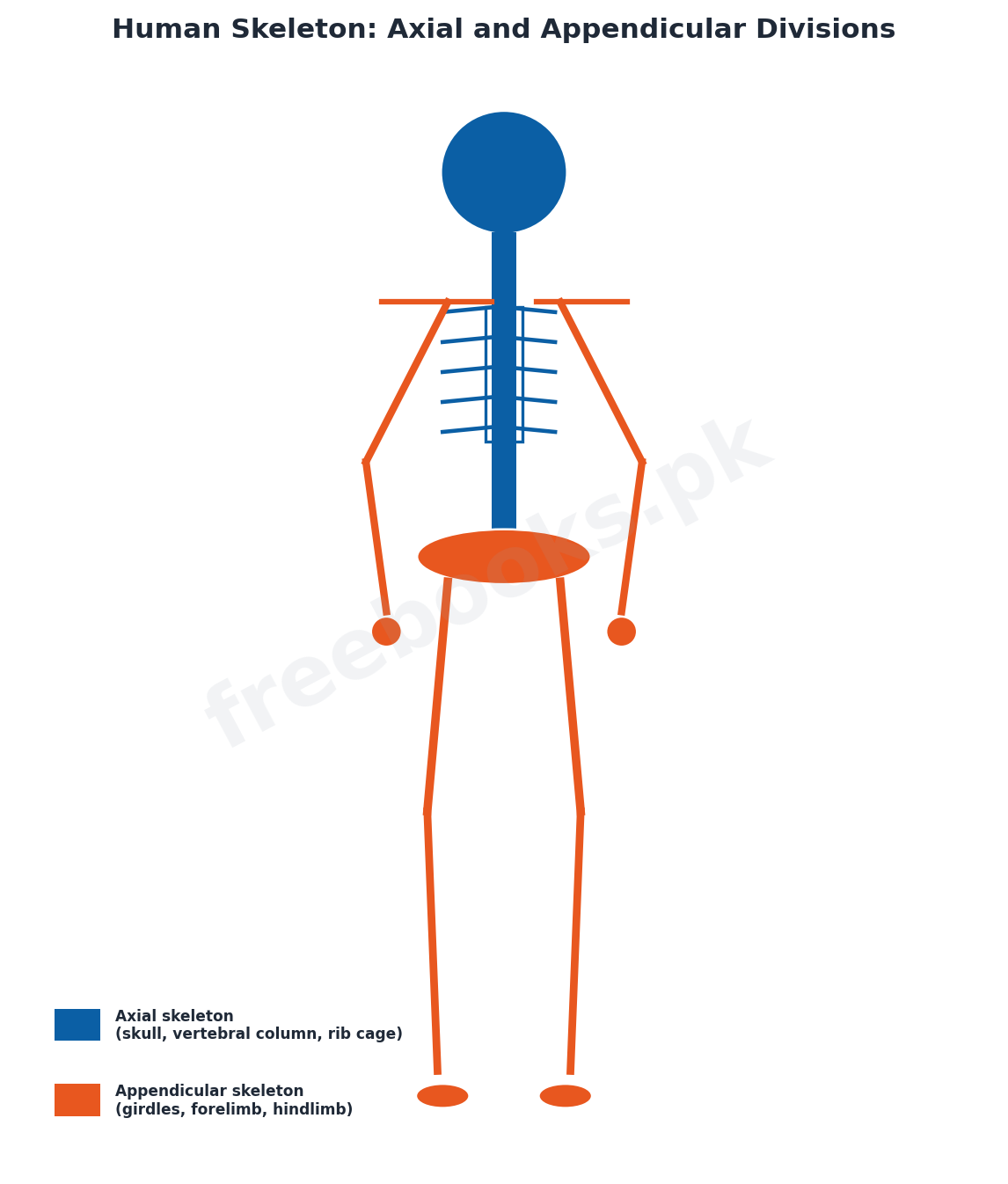
The human skeleton is divided into the axial skeleton (skull, vertebral column, ribs and sternum, forming the body's central axis) and the appendicular skeleton (the limbs and their girdles). The skull consists of the cranium (8 bones – 4 unpaired: frontal, occipital, sphenoid and ethmoid; and 2 paired: parietal and temporal – protecting the brain) and 14 facial bones (6 paired: maxilla, zygomatic, nasal, lacrimal, palatine and inferior concha; and 2 unpaired: mandible and vomer). The vertebral column, extending from skull to pelvis and protecting the spinal cord, consists of 33 vertebrae grouped as 7 cervical (neck, including the atlas and axis), 12 thoracic, 5 lumbar, and 9 pelvic vertebrae fused into the sacrum (5) and coccyx (4); its natural curvatures give it more strength than a straight column would have. The rib cage comprises 12 pairs of ribs, of which 10 pairs connect to the sternum (directly or via costal cartilage) and the lower 2 pairs are 'floating ribs' with no sternal attachment, together enclosing the chest cavity.
The appendicular skeleton consists of the pectoral girdle and forelimbs, and the pelvic girdle and hindlimbs. The pectoral girdle comprises the scapula and clavicle (which connects the scapula to the sternum); the forelimb consists of the humerus, radius and ulna, 8 carpals, 5 metacarpals and 14 phalanges, with the humerus forming a ball-and-socket joint with the scapula and a hinge joint with the radius and ulna at the elbow. The pelvic girdle consists of two coxal bones, each formed by fusion of the ilium, ischium and pubis, attaching the hindlimb to the vertebral column; the hindlimb consists of the femur, tibia and fibula, 7 tarsals, 5 metatarsals and 14 phalanges, with the femur forming a ball-and-socket hip joint proximally and a hinge knee joint distally.
Joints, Skeletal Deformities and Bone Repair
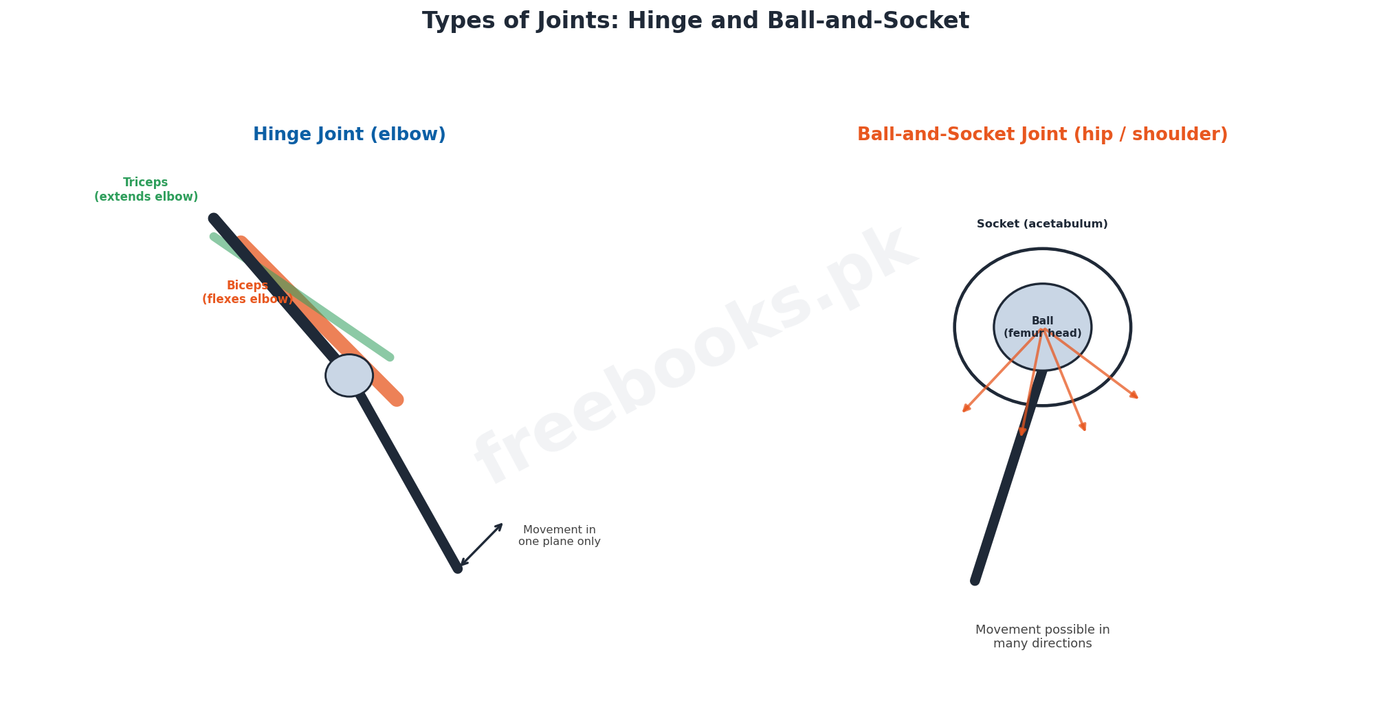
Joints, where bones meet, are classified by the movement they allow (immovable, slightly movable, or freely movable) or by structure. Fibrous joints (e.g. between skull bones) are held by short collagen fibres and allow no movement; cartilaginous joints (e.g. between vertebrae) allow little movement; and synovial joints contain a fluid-filled cavity enclosed by a fibrous capsule and synovial membrane, allowing free movement – the hinge joint (e.g. elbow, knee), permitting movement in essentially one plane, and the ball-and-socket joint (e.g. hip, shoulder), permitting movement in multiple directions, being the two major types. Skeletal deformities have varied causes: genetic causes include cleft palate and microcephaly; hormonal causes include osteoporosis (bone resorption outpacing deposition, common in postmenopausal women due to reduced estrogen); and nutritional causes include osteomalacia and rickets (softened, deformed bones from inadequate calcium or vitamin D). Other disorders include a slipped (herniated) intervertebral disc, in which the annulus fibrosus ruptures and the nucleus pulposus protrudes, potentially compressing the spinal cord or nerves; sciatica, stabbing pain from sciatic nerve injury; and arthritis, inflammatory or degenerative joint disease including osteoarthritis, rheumatoid arthritis and gouty arthritis.
A broken bone is treated by reduction (closed, manually realigning the bone; or open, surgically securing it with pins or wires) followed by immobilisation in a cast, with healing typically taking 8 to 12 weeks. Repair proceeds through four phases: hematoma formation, in which torn blood vessels form a clot at the fracture site and nearby bone cells begin to die; soft callus formation (3 to 4 weeks), in which capillaries and fibroblasts/osteoblasts invade the hematoma and begin building new bone; bony callus formation, in which osteoblasts and osteoclasts convert the soft callus into a firm bony union over 2 to 3 months; and remodeling, in which excess material is removed over several months until the repaired bone closely resembles the original structure.
Muscle Types and the Structure of Skeletal Muscle
Vertebrates possess three kinds of muscle cells. Smooth muscle is unstriated, spindle-shaped with a single nucleus, involuntary, and found in the walls of blood vessels and the digestive tract. Cardiac muscle, found only in the heart, is striated but involuntary, made of branched, interconnected cells each with its own nucleus. Skeletal muscle is striated (alternating light and dark bands) and voluntary, attached to bone via tendons, and responsible for moving the skeleton. Each skeletal muscle fibre is a large, multinucleated cylindrical cell bounded by the sarcolemma, containing sarcoplasm rich in glycogen and the oxygen-storing pigment myoglobin, and packed with parallel myofibrils built from repeating contractile units called sarcomeres. Along each myofibril, the dark A band (containing thick filaments) alternates with the light I band (containing only thin filaments, bisected by the Z line); the lighter H zone in the middle of the A band is bisected by the M line, and a sarcomere is defined as the region between two successive Z lines.
Each myofibril's myofilaments are of two types. Thick filaments (about 16 nm in diameter) are composed of myosin, a protein whose tail terminates in two globular heads, called cross bridges, which link to the thin filaments during contraction. Thin filaments (7 to 8 nm in diameter) are composed chiefly of two twisted chains of actin, wound around by two strands of tropomyosin, with the regulatory protein troponin (which binds actin, tropomyosin and calcium ions respectively via its three subunits) positioned at intervals along the filament. Each myosin filament is surrounded by six actin filaments, and it is the precisely ordered, overlapping arrangement of these thick and thin filaments that gives skeletal muscle its striped, striated appearance under the microscope.
The Sliding Filament Model and Control of Muscle Contraction
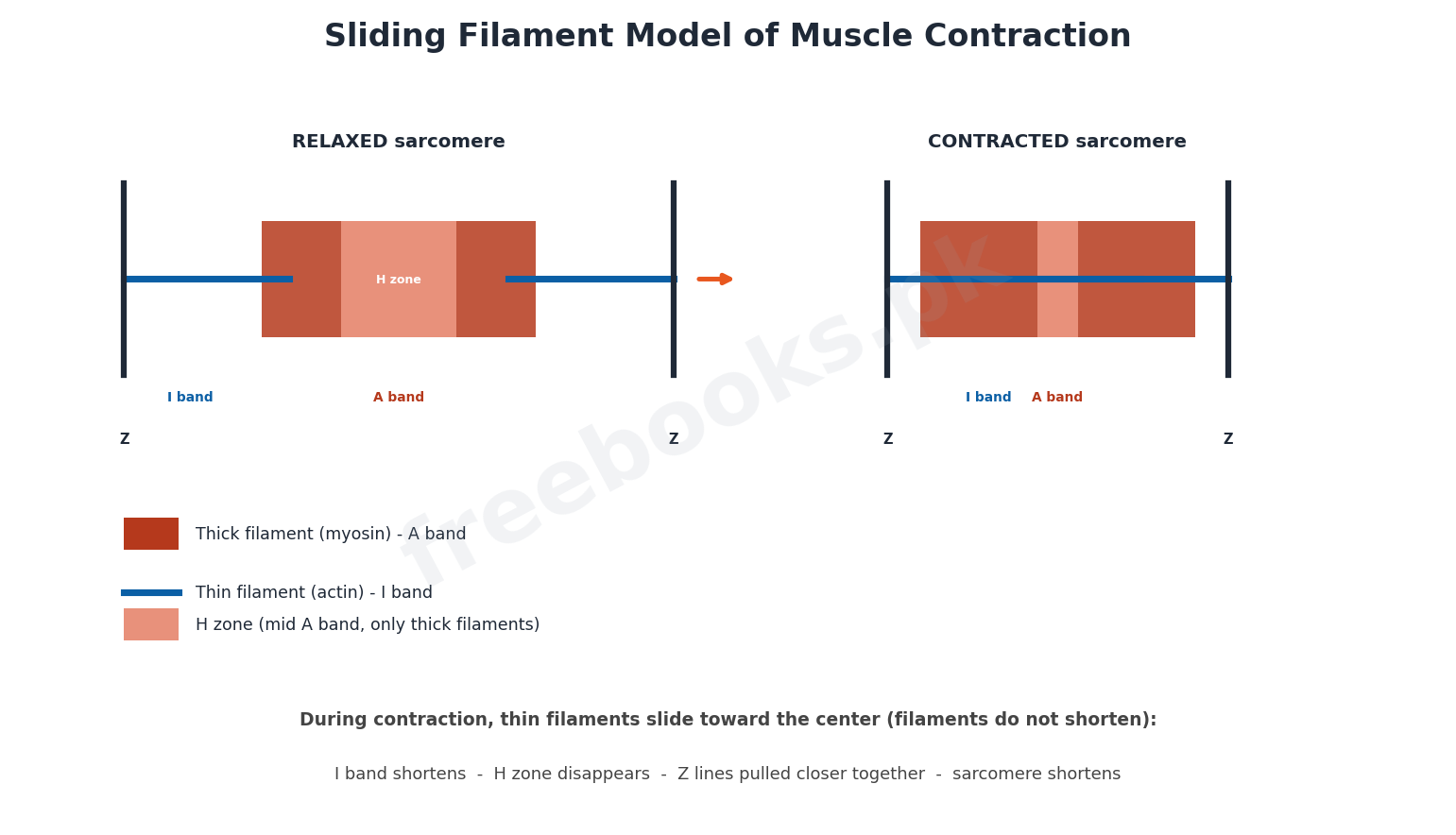
The sliding filament model, proposed by Huxley and Huxley in 1954, explains muscle contraction as the thin (actin) filaments sliding past the thick (myosin) filaments without either filament changing length, so the zones of overlap increase: the I band shortens, the H zone disappears, and successive Z lines are pulled closer together, shortening the whole sarcomere and hence the whole muscle fibre. In this process, the myosin cross bridges repeatedly attach to specific binding sites on the actin filament, bend to pull the actin filament toward the centre of the sarcomere, detach (using ATP hydrolysis), and reattach further along, in a repeating cycle. At rest, tropomyosin physically blocks these myosin-binding sites on actin, preventing contraction.
Muscle contraction is triggered by a nerve impulse at the neuromuscular junction, which travels deep into the muscle fibre via T-tubules to the adjacent sarcoplasmic reticulum, releasing calcium ions into the cytosol. These calcium ions bind to troponin, causing a conformational shift that displaces tropomyosin and exposes the myosin-binding sites on actin, allowing cross bridges to form and the sliding filament cycle to proceed – each fibre contracts according to an 'all or none' principle, with the overall strength of a muscle's contraction depending on how many of its fibres are simultaneously activated. ATP is essential not just for the power stroke but also to break the link between myosin and actin at the end of each cycle; after death, falling ATP levels leave the cross bridges permanently locked, causing the muscle stiffness known as rigor mortis. Ongoing ATP supply during life comes primarily from the aerobic breakdown of glucose (from stored glycogen) and creatine phosphate, with anaerobic glycolysis (producing lactic acid) supplementing this during oxygen deficiency or intense activity; excess lactic acid buildup contributes to muscle fatigue, a state of physiological inability to contract effectively, distinct from tetany (muscle twitching and spasm from low blood calcium) and cramp (a sudden, painful, sustained involuntary contraction).
Muscle Arrangement and Locomotion in Invertebrates
A skeletal muscle has three functional parts: the origin, the fixed end that does not move when the muscle contracts; the insertion, the end attached to the bone that is moved; and the belly, the thick, contractile middle portion. Muscles connect to bones via non-elastic tendons, while ligaments (slightly elastic) connect bone to bone across a joint. Because a single muscle can only pull, never push, movable joints depend on antagonistic muscle pairs, in which one muscle's contraction is reversed by the contraction of its partner – the classic example being the biceps brachii (which bends, or flexes, the arm at the elbow) and the triceps (which straightens, or extends, it), which never contract simultaneously.
Invertebrates and protoctists show a remarkable diversity of locomotory mechanisms. Euglena moves by lashing an anterior flagellum, whose spiral, whip-like beating pulls the organism forward, aided by contractile myonemes that allow direction change (Euglenoid movement). Paramecium moves by the coordinated, wave-like beating of many short cilia, each performing an effective stroke (rapid bend) followed by a recovery stroke (straightening), powered by ATP. Amoeba moves by extending finger-like pseudopodia in the direction of cytoplasmic flow, causing the whole cell body to follow. Jellyfish use jet propulsion, drawing water into their bell-shaped body and then forcefully expelling it to drive themselves forward. The earthworm uses an accordion-like peristaltic movement, alternating contraction of circular muscles (which elongates and thins segments) and longitudinal muscles (which shortens and thickens segments), anchored at each stage by setae. The cockroach walks using alternating tripod support from its six legs and can also fly using its posterior wing pair, while the starfish moves using water-powered tube feet arranged along each arm, extending, attaching by suction, and then contracting to pull the body along.
Locomotion in Vertebrates
Vertebrate locomotion shows adaptations specific to each medium and habitat. Fish are propelled by muscular contractions passing along the body from anterior to posterior, producing S-shaped lashing movements; their streamlined, mucus-coated bodies minimise friction, with unpaired dorsal and ventral fins providing stability, paired pectoral and pelvic fins used for steering, the caudal fin providing forward thrust, and a swim bladder maintaining buoyancy in bony fish. Amphibians combine a fish-like body with limbs, either wriggling on the belly using segmental muscles or, in frogs and toads, using powerful hind limbs for swimming and jumping. Reptiles show a more ossified, better-supported skeleton adapted for walking and running, with a modified atlas-axis complex allowing greater head rotation; some prehistoric reptiles were bipedal, freeing the front limbs for prey capture or eventually flight.
Birds have a skeleton highly modified for flight: bones with large air spaces reduce weight, the sternum is expanded into a keel for the attachment of powerful flight muscles, and the body is covered in feathers that both provide lift surface and insulation. Flight may be passive (gliding, using the wing as an aerofoil to generate lift from airflow) or active (flapping, in which faster airflow over the curved upper wing surface than the lower surface creates a net upward lifting pressure). Mammals show three modes of limb-based locomotion: plantigrade (walking flat on the sole, as in humans and bears), digitigrade (walking on the digits, faster, as in dogs and rodents), and unguligrade (walking on hoof-modified toe tips, the fastest, as in deer and horses); in running mammals, the spine itself flexes and extends with each stride to increase stride length and power. Across land vertebrates (tetrapods), a common pentadactyl limb plan and a firmly sacrum-attached pelvic girdle (built from the ilium, ischium and pubis meeting at the acetabulum, which articulates with the femur) underlie the diverse locomotory adaptations seen from walking and running to the independently evolved flight of pterodactyls, birds and bats.
Important Definitions
Turgor pressure
The internal hydrostatic pressure that develops in a plant cell as water enters the vacuole by osmosis, keeping the cell rigid and resistant to bending.
Sclerenchyma
Plant support tissue composed of cells with thick, lignin-impregnated secondary walls, mostly non-living, including fibres, sclereids and vessels.
Ecdysis
The periodic shedding and replacement of the exoskeleton in arthropods, allowing growth, controlled by the hormone ecdysone.
Sarcomere
The region of a myofibril between two successive Z lines, and the smallest contractile unit of a skeletal muscle fibre.
Antagonism
The relationship between a pair of muscles at a joint in which the contraction of one reverses the action of the other, such as the biceps and triceps.
Tropism
A directional growth movement of a plant organ toward or away from an external stimulus such as light, gravity or touch.
Nastic movement
A non-directional movement of a plant part in response to an external stimulus, such as the opening and closing of a flower with light or temperature.
Rigor mortis
The stiffening of the body after death caused by falling ATP levels, which leaves myosin-actin cross bridges permanently locked in place.
Key Facts
| Item | Fact |
|---|---|
| Cranium bones | 8 cranial bones (4 unpaired: frontal, occipital, sphenoid, ethmoid; 2 paired: parietal, temporal). |
| Vertebral column | 33 vertebrae: 7 cervical, 12 thoracic, 5 lumbar, and 9 pelvic (5 fused as sacrum, 4 fused as coccyx). |
| Rib pairs | 12 pairs of ribs; 10 pairs attach to the sternum, the lower 2 pairs are floating ribs. |
| Forelimb bones | Humerus, radius, ulna, 8 carpals, 5 metacarpals, 14 phalanges. |
| Hindlimb bones | Femur, tibia, fibula, 7 tarsals, 5 metatarsals, 14 phalanges. |
| Total skeletal muscles | About 650 muscles occur in the human body, most arranged in antagonistic pairs. |
| Filament diameters | Thick (myosin) filaments ~16 nm diameter; thin (actin) filaments ~7-8 nm diameter. |
| Bone healing time | Simple fractures typically heal in about 8-12 weeks, longer in elderly patients. |
Diagrams & Illustrations
Sliding Filament Model of Muscle Contraction: a diagram of a sarcomere at rest and during contraction, showing the Z lines, A band, I band and H zone, and the thick (myosin) and thin (actin) filaments sliding past each other to shorten the sarcomere.
Human Skeleton: Axial and Appendicular Divisions: a labelled diagram of the human skeleton with the axial skeleton (skull, vertebral column, rib cage) and appendicular skeleton (pectoral girdle, forelimb, pelvic girdle, hindlimb) shown in different colours.
Types of Joints: Hinge and Ball-and-Socket: a comparison diagram of a hinge joint (elbow, with biceps and triceps as an antagonistic pair) and a ball-and-socket joint (hip or shoulder, showing its wider range of movement).
Short Questions & Answers
Differentiate sclerenchyma and collenchyma cells.
Sclerenchyma cells have thick, lignified secondary walls, are mostly non-living, and provide rigid support to mature plant parts; collenchyma cells have only angular thickening of their primary walls, remain living, and provide flexible support to young, growing herbaceous parts.
What is ecdysis, and why is it necessary?
Ecdysis is the periodic shedding and replacement of the exoskeleton in arthropods, controlled by the hormone ecdysone; it is necessary because the hardened, non-living exoskeleton cannot grow, so it must be shed and replaced with a larger one to allow the animal to increase in size.
Differentiate a hinge joint and a ball-and-socket joint, with one example each.
A hinge joint allows movement in essentially one plane, such as at the elbow or knee; a ball-and-socket joint allows movement in multiple directions and planes, providing much greater flexibility, such as at the hip or shoulder.
What is the sliding filament model of muscle contraction?
The sliding filament model states that during muscle contraction, the thin (actin) filaments slide past the thick (myosin) filaments without either filament shortening, increasing their overlap, which shortens the I band, causes the H zone to disappear, and pulls successive Z lines closer together, shortening the sarcomere.
What role does calcium play in muscle contraction?
Calcium ions released from the sarcoplasmic reticulum bind to troponin on the thin filament, causing a shift that displaces tropomyosin and exposes the myosin-binding sites on actin, allowing cross bridges to form and the contraction cycle to proceed.
Differentiate plantigrade and digitigrade locomotion, with an example of each.
In plantigrade locomotion, the sole, wrist/heel and digits all rest on the ground while walking, as in humans and bears; in digitigrade locomotion, the animal walks on its digits only (with the first digit often reduced or lost), allowing faster movement, as in dogs and rodents.
Long Questions & Answers
Describe the three main types of skeleton found in animals, with examples.
Animals depend on one of three basic types of skeletal support. A hydrostatic skeleton relies on a fluid-filled body cavity against which surrounding muscles can contract, converting muscular force into changes in body shape and length without any rigid skeletal elements; it is found in cnidarians such as the sea anemone, whose contraction of circular muscles pressurises its water-filled cavity to maintain an upright form, and in annelids such as the earthworm, whose fluid-filled compartments (separated by septa) allow circular muscle contraction to elongate a segment and longitudinal muscle contraction to shorten it, producing the alternating waves of elongation and contraction (aided by setae) that move the animal through soil. An exoskeleton is a hardened, non-living covering secreted externally by the ectoderm, consisting of an outer waterproof epicuticle and a thicker procuticle made of chitin and protein, further toughened by sclerotization or, in some molluscs, calcium carbonate; the simplest exoskeletons are seen in molluscs such as snails and bivalves, while arthropods (such as insects and crustaceans) have the most elaborate exoskeletons, modified with flexible joints for movement, sensory bristles, and structures permitting gas exchange, though because this rigid, non-living covering cannot grow with the animal, arthropods must periodically undergo ecdysis – shedding the old exoskeleton and secreting a larger new one – a hormonally controlled process during which the animal is briefly vulnerable to predators. An endoskeleton, found in vertebrates, is an internal framework built of living connective tissues, chiefly bone (rigid, hardened by calcium phosphate deposits in a collagen matrix, with a dense compact outer layer and a lighter, porous spongy interior housing blood-cell-producing marrow) and cartilage (softer, avascular, and secreted by chondrocytes into a flexible collagen matrix, found at joints and in structures such as the outer ear); because an endoskeleton grows along with the animal's own tissues, it does not impose the same size restriction that an exoskeleton does, and it also provides an internal framework for muscle attachment that can support the greater size typical of many vertebrates.
Describe the structure of skeletal muscle from the whole muscle down to the level of the myofilaments.
A skeletal muscle is composed of bundles of individual muscle fibres, each of which is a single, remarkably large, multinucleated cylindrical cell, typically 10 to 100 micrometres in diameter, bounded by a specialised cell membrane called the sarcolemma; its cytoplasm, the sarcoplasm, is distinguished from that of ordinary cells by large stores of glycogen and by myoglobin, a red, oxygen-binding pigment that supplies oxygen to the fibre's numerous mitochondria. Within each fibre, running the entire length of the cell in parallel array, are large numbers of thread-like myofibrils, each 1 to 2 micrometres in diameter, and it is these myofibrils, viewed under high magnification, that reveal the alternating dark and light banding responsible for skeletal muscle's characteristic striped appearance: the dark A bands (so named because they are anisotropic, or capable of polarising light) alternate with the light I bands (isotropic, non-polarising); within each A band is a lighter central H zone, itself bisected by a dark M line, while each I band is bisected by a dense Z line. The region of a myofibril lying between two successive Z lines is called a sarcomere, the fundamental repeating contractile unit of the entire muscle fibre, and each sarcomere is built from two types of myofilaments arranged with remarkable precision: thick filaments, about 16 nanometres in diameter and composed of the protein myosin – each myosin molecule having a long tail of two coiled polypeptide chains terminating in two globular heads, or cross bridges, that project outward to interact with neighbouring thin filaments – occupy the entire length of the A band; and thin filaments, only 7 to 8 nanometres in diameter and composed chiefly of two twisted chains of the globular protein actin, wound about by two helical strands of the regulatory protein tropomyosin, with the three-part regulatory protein complex troponin (one subunit binding actin, one binding tropomyosin, and one binding calcium ions) positioned at intervals along its length, extend across the I band and partway into the neighbouring A bands, with each thick filament typically surrounded by six thin filaments in the region of overlap. It is the sliding of these two types of interdigitated filaments past one another, driven by the repeated attachment, power-stroke and detachment cycling of the myosin cross bridges against binding sites on actin (a cycle regulated by calcium ions displacing tropomyosin, and powered by ATP hydrolysis), that produces the shortening of each sarcomere and, multiplied across millions of sarcomeres in series and parallel throughout the muscle, the visible contraction and force generation of the whole muscle.
Explain the sliding filament model of muscle contraction and describe how calcium ions and ATP regulate the process.
The sliding filament model, put forward by Huxley and Huxley in 1954, remains the accepted explanation of how skeletal muscle shortens during contraction. According to this model, neither the thick (myosin) filaments nor the thin (actin) filaments of a sarcomere change in length during contraction; instead, the thin filaments are actively pulled inward, sliding past the stationary thick filaments so that the two sets of filaments come to overlap over a greater proportion of their length. This increasing overlap has several visible consequences at the level of the sarcomere: the I band, which contains only thin filaments, progressively shortens as thin filaments slide further into the A band region; the H zone, the central region of the A band that normally contains no overlapping thin filaments, narrows and eventually disappears as thin filaments from either side meet in the middle; and, because both I bands attached to a given A band are shortening symmetrically, the two Z lines bounding that sarcomere are drawn closer together, shortening the sarcomere as a whole, and this shortening, repeated over many thousands of sarcomeres arranged end to end along each myofibril, produces the visible shortening of the entire muscle fibre and ultimately the whole muscle. This filament sliding is driven directly by the myosin cross bridges, the globular heads projecting from the thick filament, which cyclically attach to specific binding sites on the adjacent actin filament, bend (the power stroke) to drag the thin filament a short distance toward the centre of the sarcomere, detach, and then reattach further along the actin filament to repeat the cycle – but this cycle can only proceed if the myosin-binding sites on actin are physically accessible, and at rest they are not, being blocked by the regulatory protein tropomyosin, which winds along the actin filament precisely covering these sites. The trigger that removes this block is a nerve impulse arriving at the neuromuscular junction, which is conducted deep into the muscle fibre via the T-tubule system to the closely associated sarcoplasmic reticulum, causing it to release stored calcium ions into the surrounding cytosol; these calcium ions bind to troponin, the regulatory protein complex positioned along the thin filament, causing a conformational change that physically displaces tropomyosin away from the myosin-binding sites on actin, at last permitting the cross bridges to attach and the sliding cycle to begin. ATP is essential to this process at two distinct points: it powers the myosin cross bridge's ability to detach from actin after each power stroke, resetting it to bind further along the filament and repeat the cycle, and it is also required to actively pump calcium ions back into the sarcoplasmic reticulum once the stimulating nerve impulse ceases, allowing tropomyosin to re-cover the binding sites and the muscle to relax; when ATP becomes unavailable after death, cross bridges that have already attached to actin cannot detach and so remain permanently locked in place, producing the total-body muscular stiffness known as rigor mortis.
Describe the axial and appendicular divisions of the human skeleton in detail.
The human skeleton is organised into two major divisions, the axial skeleton and the appendicular skeleton, which together provide the body's complete bony framework. The axial skeleton forms the central, vertical axis of the body and consists of the skull, vertebral column, ribs and sternum. The skull itself is composed of two sets of bones: the cranium, made up of eight bones – four unpaired (the frontal, occipital, sphenoid and ethmoid bones) and two paired (the parietal and temporal bones) – which together enclose and protect the brain, and the facial skeleton, made up of fourteen bones, six paired (the maxilla, zygomatic, nasal, lacrimal, palatine and inferior concha bones) and two unpaired (the mandible, the only movable skull bone, and the vomer). Below the skull, the vertebral column extends down to the pelvis, encasing and protecting the delicate spinal cord within its central canal, and is built from thirty-three individual vertebrae grouped into four named regions – seven cervical vertebrae in the neck (the first two of which, the atlas and axis, are specially modified to support and rotate the head), twelve thoracic vertebrae in the upper back (each articulating with a pair of ribs), five lumbar vertebrae in the lower back, and nine pelvic vertebrae that fuse during development into two solid structures, the sacrum (from five vertebrae) and the coccyx, or tailbone (from four vertebrae) – with the column's natural forward-and-backward curvatures giving it substantially greater mechanical strength than a perfectly straight column of the same material would have. Completing the axial skeleton, the rib cage consists of twelve pairs of ribs that articulate posteriorly with the thoracic vertebrae; anteriorly, the upper ten pairs connect, directly or via a shared costal cartilage arch, to the flat sternum in the midline of the chest, while the lowest two pairs, lacking any sternal attachment, are termed floating ribs, and together this bony cage encloses and protects the semi-vacuum chest cavity housing the heart and lungs. The appendicular skeleton, by contrast, consists of the bones of the limbs and the girdles that attach them to the axial skeleton. The pectoral girdle, comprising the scapula (shoulder blade) and clavicle (collarbone, which anchors the scapula to the sternum), attaches the forelimb, which itself consists of a single long humerus (forming a highly mobile ball-and-socket joint with the scapula at the shoulder, and a hinge joint with the forearm bones at the elbow), the paired radius and ulna of the forearm, eight small wrist bones called carpals, five metacarpals forming the framework of the palm, and fourteen phalanges arranged in five digits. The pelvic girdle, consisting of two coxal bones (each itself formed by the fusion of three separate bones, the ilium, ischium and pubis), firmly attaches the hindlimb to the base of the vertebral column and supports the weight of the pelvic region; the hindlimb consists of a single femur (forming a ball-and-socket hip joint proximally with the coxal bone, and a hinge knee joint distally with the shin bones), the paired tibia and fibula of the lower leg, seven tarsal bones of the ankle, five metatarsals forming the framework of the foot, and fourteen phalanges of the toes, mirroring the structural plan of the forelimb in accordance with the shared pentadactyl body plan of tetrapod vertebrates.
Compare locomotion in fish, birds and mammals, highlighting the key adaptations of each group.
Locomotion in vertebrates has been shaped by the very different physical demands of moving through water, air and on land, producing three strikingly different sets of adaptations in fish, birds and mammals. Fish are propelled almost entirely by muscular contractions that pass rhythmically along the body from anterior to posterior, with alternating contraction of muscle blocks on either side of the body producing a characteristic lashing, S-shaped wave of motion that drives the animal forward, a pattern especially clear in cartilaginous fish such as sharks and dogfish; this propulsive mechanism is supported by a strongly streamlined, tapered body shape that minimises drag as water flows over it, further aided by mucus or oily secretions that lubricate the body surface (over scales in bony fish, or dermal denticles in cartilaginous fish) to reduce friction still further, while the fins serve distinct stabilising and steering roles – the unpaired dorsal and ventral fins resist rolling and yawing to keep the fish stable, the paired pectoral and pelvic fins are used for fine steering, balance and braking, and the caudal (tail) fin, working with body undulations, provides the main forward thrust – and buoyancy in bony fish is finely regulated by an internal gas-filled swim bladder, allowing the fish to remain suspended at a chosen depth with minimal muscular effort. Birds, by contrast, are adapted for locomotion through air, an environment that offers little support and requires the generation of active lift; their skeleton is extensively lightened by bones containing large internal air spaces, while their forelimbs have evolved into wings and their sternum has been dramatically enlarged and keeled to provide a greatly expanded surface area for the attachment of the massive pectoral flight muscles needed to power the wingbeat, and the body is covered by feathers that both enlarge the aerodynamic surface of the wing and provide the insulation needed to sustain the very high metabolic rate flight demands; birds achieve flight either passively, by gliding with wings held as aerofoils to extract lift from oncoming or rising air currents, or actively, by flapping the wings so that air is forced to flow faster over their curved upper surface than beneath them, lowering pressure above the wing relative to below it and so generating a continuous net upward lift force. Mammals, finally, remain fundamentally adapted for locomotion on solid ground (with notable secondary adaptations to water, air and burrowing in particular lineages), and show a graded series of limb postures reflecting different balances of stability versus speed: plantigrade mammals such as humans and bears place the entire sole of the foot, including wrist or heel and digits, flat on the ground with each step, a stable but comparatively slow gait; digitigrade mammals such as dogs and rodents walk balanced on their digits alone, with the first digit often reduced or absent, gaining speed at some cost to stability; and unguligrade mammals such as deer and horses walk on the very tips of their toes, which have become modified into hardened hooves, representing the fastest and most specialised of the three gaits; in fast-running mammals generally, the vertebral column itself flexes and extends with each stride, working in concert with limb muscles to lengthen stride and add propulsive power well beyond what the limbs alone could generate.
MCQs with Answers
The internal hydrostatic pressure that keeps parenchyma cells rigid is called: (a) osmotic pressure (b) turgor pressure (c) root pressure (d) wall pressure
Correct Answer: (b) turgor pressure. Turgor pressure develops as water enters the vacuole by osmosis, keeping parenchyma cells rigid and resistant to bending.
The rapid folding of Mimosa leaflets on touch is an example of which type of movement? (a) tropic movement (b) nastic (turgor) movement (c) phototactic movement (d) chemotropic movement
Correct Answer: (b) nastic (turgor) movement. This is a turgor movement, a type of autonomic movement caused by rapid loss of turgor pressure in pulvinus cells.
Ecdysis, the periodic shedding of the exoskeleton, is controlled by the hormone: (a) insulin (b) ecdysone (c) auxin (d) aldosterone
Correct Answer: (b) ecdysone. Ecdysone controls the hormonally regulated process of ecdysis (molting) in arthropods.
The human cranium is made up of how many bones? (a) 6 (b) 8 (c) 14 (d) 22
Correct Answer: (b) 8. The cranium consists of 8 bones: 4 unpaired (frontal, occipital, sphenoid, ethmoid) and 2 paired (parietal, temporal).
A joint that allows movement in several directions, such as the hip or shoulder, is a/an: (a) hinge joint (b) fibrous joint (c) ball-and-socket joint (d) cartilaginous joint
Correct Answer: (c) ball-and-socket joint. Ball-and-socket joints, found at the hip and shoulder, allow movement in multiple directions and provide maximum flexibility.
In a sarcomere, the zone containing only thin (actin) filaments is the: (a) A band (b) H zone (c) I band (d) M line
Correct Answer: (c) I band. The I band is the light, isotropic zone containing only thin filaments, bisected by the Z line.
According to the sliding filament model, during contraction: (a) thick and thin filaments both shorten (b) thin filaments slide past thick filaments without either shortening (c) only the thick filaments move (d) the Z lines move further apart
Correct Answer: (b) thin filaments slide past thick filaments without either shortening. In the sliding filament model, filaments do not shorten; thin filaments slide past thick filaments, pulling Z lines closer together.
The muscle protein that blocks the myosin-binding sites on actin at rest is: (a) troponin (b) tropomyosin (c) myosin (d) actin
Correct Answer: (b) tropomyosin. Tropomyosin covers the myosin-binding sites on actin at rest; it is displaced when calcium binds troponin.
Stiffening of the body after death, caused by depleted ATP locking cross bridges in place, is called: (a) tetany (b) rigor mortis (c) muscle fatigue (d) cramp
Correct Answer: (b) rigor mortis. Rigor mortis occurs because falling ATP levels after death prevent myosin cross bridges from detaching from actin.
Mammals that walk on hoof-modified toe tips, such as deer, show which type of locomotion? (a) plantigrade (b) digitigrade (c) unguligrade (d) hydrostatic
Correct Answer: (c) unguligrade. Unguligrade locomotion, walking on hoof-modified toe tips, is the fastest of the three mammalian limb postures.
Quick Revision Summary
- Plant support: parenchyma (turgor pressure), sclerenchyma (lignified, rigid, non-living: fibres/sclereids/vessels), collenchyma (living, flexible, young parts). Secondary growth: vascular cambium (secondary xylem/phloem) + cork cambium -> growth rings, sapwood vs heartwood.
- Plant movement: Autonomic (tactic, turgor, growth movements – internal cause) vs Paratonic (tropic, nastic – external cause). Hormones: auxin (phototropism/gravitropism), abscisic acid vs gibberellins (nastic balance).
- Animal skeletons: Hydrostatic (fluid cavity + muscle, e.g. earthworm) -> Exoskeleton (chitin, arthropods, needs ecdysis via ecdysone) -> Endoskeleton (bone + cartilage, vertebrates; osteoblast/osteocyte/osteoclast).
- Human skeleton: Axial (skull: 8 cranial + 14 facial bones; vertebral column: 33 vertebrae, 7C+12T+5L+9 pelvic; ribs: 12 pairs, 2 floating) + Appendicular (pectoral girdle + forelimb: humerus/radius/ulna/8 carpals/5 metacarpals/14 phalanges; pelvic girdle + hindlimb: femur/tibia/fibula/7 tarsals/5 metatarsals/14 phalanges).
- Joints: fibrous (immovable) / cartilaginous (slight) / synovial (free: hinge = one plane, e.g. elbow; ball-and-socket = multi-directional, e.g. hip). Deformities: genetic (cleft palate), hormonal (osteoporosis – low estrogen), nutritional (osteomalacia/rickets – low Ca/vitamin D). Fracture repair: hematoma -> soft callus -> bony callus -> remodeling (8-12 weeks).
- Muscle types: smooth (unstriated, involuntary), cardiac (striated, involuntary), skeletal (striated, voluntary). Sarcomere = Z line to Z line; A band (myosin) + I band (actin, has Z line) + H zone (mid A band). Sliding filament model (Huxley, 1954): actin slides on myosin -> I band and H zone shrink, sarcomere shortens.
- Contraction control: nerve impulse -> T-tubules -> SR releases Ca2+ -> Ca2+ binds troponin -> tropomyosin shifts -> cross bridges form -> ATP powers detachment/reattachment. No ATP after death = rigor mortis. Energy: aerobic glycolysis + creatine phosphate, anaerobic (lactic acid) under stress -> fatigue. Locomotion: Euglena (flagellum), Paramecium (cilia), Amoeba (pseudopodia), earthworm (peristalsis+setae), fish (S-wave+fins), birds (keel+flapping/gliding), mammals (plantigrade/digitigrade/unguligrade). Notes by freebooks.pk.
Exam Tips
- Build a simple 2-column table of autonomic vs paratonic plant movements with one named example in each sub-category (tactic/turgor/growth vs tropic/nastic) – this structure matches how these questions are usually marked.
- Memorise the exact bone counts for the human skeleton (8 cranial, 14 facial, 33 vertebrae split 7-12-5-9, 12 rib pairs) – fill-in-the-blank and short questions frequently target these numbers directly.
- Learn the sarcomere diagram (Z line, I band, A band, H zone, M line) well enough to redraw it from memory in both the relaxed and contracted state, showing exactly which zones shrink or disappear.
- Walk through muscle contraction as one continuous numbered sequence (nerve impulse -> T-tubule -> Ca2+ release -> troponin -> tropomyosin shift -> cross bridge cycling -> ATP-dependent detachment) rather than memorising isolated facts – long questions are marked on this sequence.
- Keep hinge joint and ball-and-socket joint clearly separated with one labelled example each, and be ready to explain antagonistic muscle pairs (biceps/triceps) as the mechanism that moves a hinge joint in both directions.
- Group locomotion examples by mechanism, not just by animal name: flagellar (Euglena), ciliary (Paramecium), pseudopodial (Amoeba), jet propulsion (jellyfish), peristaltic (earthworm), tube feet (starfish) – this is a very common matching-style question.
- For plantigrade/digitigrade/unguligrade, remember the speed order (plantigrade slowest, unguligrade fastest) together with one clear example of each so you never confuse the three.