This chapter covers Chromosomes and DNA from the 2nd Year (FSc Part-II) Biology syllabus of the Punjab Curriculum and Textbook Board (PTB/PCTB). Chromosomes are thread-like structures that appear inside the nucleus at cell division, first observed by Walther Flemming in 1882; found in all eukaryotic cells, their number varies widely between species (humans have 46, in 23 pairs), and each chromosome carries hundreds or thousands of genes essential for normal development and survival. These notes are prepared by freebooks.pk.
The chapter covers chromosome structure and composition, the chromosomal theory of inheritance (Sutton, Morgan), the classic experiments proving DNA is the hereditary material (Griffith, Avery-MacLeod-McCarty, Hershey-Chase), the chemical nature and double helical structure of DNA (Levene, Chargaff, Franklin, Watson and Crick), semi-conservative DNA replication (Meselson-Stahl) and the replication process, the concept of the gene (Garrod, Beadle and Tatum's one gene/one polypeptide hypothesis), the central dogma of gene expression (transcription, the genetic code, and translation), and mutations.
Learning Objectives
- Describe the structure, composition and types of chromosomes, and explain karyotype.
- Describe the chromosomal theory of inheritance and the evidence provided by Sutton and Morgan.
- Describe the classic experiments (Griffith, Avery, Hershey-Chase) that established DNA as the hereditary material.
- Describe the chemical composition and double helical structure of DNA (Levene, Chargaff, Franklin, Watson and Crick).
- Explain semi-conservative DNA replication and the Meselson-Stahl experiment, and describe the replication process.
- Explain the concept of a gene using Garrod's and Beadle and Tatum's work (one gene/one polypeptide hypothesis).
- Describe the central dogma of gene expression: transcription, the genetic code, and translation.
- Describe the types and significance of mutations.
Key Concepts
Chromosome Structure, Composition and the Chromosomal Theory of Inheritance
Chromosomes are thread-like structures that appear inside the nucleus at cell division, first observed by the German embryologist Walther Flemming in 1882 in dividing salamander larvae cells; found in all eukaryotes, their number varies widely between species (Penicillium has one pair, some ferns over 500 pairs, mosquito 6, honeybee 32, corn 20, sugarcane 80, frog 26, mouse 40, and humans 46, in 23 pairs), and each carries hundreds or thousands of genes essential for normal development, so losing part or all of a chromosome has serious, often fatal, consequences. A typical chromosome consists of chromatids, a centromere (primary constriction), and a secondary constriction; chromosomes vary in size, staining properties, centromere position, and arm length, and the particular array of chromosomes an individual possesses is called its karyotype. Based on centromere position, chromosomes are classified as telocentric, acrocentric, submetacentric or metacentric, and acquire characteristic I, J or V shapes at anaphase during cell division. Chromosomes are composed of roughly 40% DNA and 60% protein, with associated RNA (since they are sites of RNA synthesis); a typical human chromosome's DNA contains about 140 million nucleotides and would measure about 5 centimetres if stretched out, yet is tightly coiled to fit inside the nucleus. Every 200 nucleotides, DNA coils around a core of eight positively charged histone proteins to form a nucleosome (resembling a string of beads), with further coiling into supercoils; permanently condensed, non-expressed chromatin is called heterochromatin, while chromatin that condenses only during cell division (and is otherwise open and expressible) is called euchromatin.
A central role for chromosomes in heredity was first suggested in 1900 by Karl Correns, and the chromosomal theory of inheritance was formulated in 1902 by Walter Sutton, based on evidence that gametes (with little cytoplasm) must carry hereditary material in their nuclei, that diploid individuals have two copies of each chromosome (matching Mendel's model of two gene copies) while gametes have one, and that chromosome pairs segregate and assort independently during meiosis, just as Mendelian traits do. A puzzle remained: organisms show far more independently assorting traits than chromosome pairs. In 1910, Thomas Hunt Morgan, studying the fruit fly Drosophila melanogaster, found a mutant white-eyed male; crossing it with a normal red-eyed female produced all red-eyed F1 offspring, but crossing F1 flies together produced F2 offspring in which all white-eyed flies were male, an unexpected result unexplained by simple Mendelian theory. A test cross of F1 females with the original white-eyed male produced a 1:1:1:1 ratio of white and red-eyed males and females, showing white-eyed females could exist; the explanation was that the white-eye gene resides only on the X chromosome (absent from Y), making it sex-linked – a trait determined by a gene on the X chromosome. This experiment provided the first clear evidence that Mendelian genes reside on chromosomes, confirming the chromosomal theory of inheritance.
DNA as Hereditary Material: Griffith, Avery, and Hershey-Chase
The first evidence of DNA's hereditary role came from British microbiologist Frederick Griffith, working with Streptococcus pneumoniae. The virulent S strain (smooth colonies, polysaccharide coat) killed infected mice, while the mutant R strain (rough colonies, no coat) did not; dead S bacteria alone were harmless, but a mixture of dead S bacteria and live, harmless R bacteria killed the mice, and live virulent S bacteria were recovered from their blood. Griffith concluded that genetic information specifying the polysaccharide coat had passed from the dead S bacteria to the living R bacteria, permanently transforming them into the virulent S form – a process he called transformation, the transfer of genetic material between cells altering the recipient's genetic makeup. The identity of the 'transforming principle' remained unknown until 1944, when Oswald Avery, Colin MacLeod and Maclyn McCarty purified Griffith's transforming material to 99.98% protein-free purity without any loss of transforming activity; protein- and RNA-digesting enzymes had no effect on this activity, but the DNA-digesting enzyme DNase destroyed it completely, identifying DNA as the transforming principle.
Further confirmation came in 1952 from Alfred Hershey and Martha Chase, working with bacteriophage T2 viruses. They labelled viral DNA with radioactive phosphorus (32P) in one experiment and viral protein coats with radioactive sulphur (35S) in another; after infecting bacteria and violently agitating the mixture to strip off the empty protein coats, nearly all the 35S label was removed from the bacteria, while the 32P label had entered the bacterial cells and was later found in the next generation of viruses. This proved that the hereditary information transmitted into the bacteria to direct new virus production was DNA, not protein, providing strong independent confirmation of Avery's earlier conclusion.
Chemical Nature and Double Helical Structure of DNA
DNA was first discovered in 1869 by German chemist Friedrich Miescher, who extracted a substance he called 'nuclein' (later 'nucleic acid') from cell nuclei and fish sperm; its function remained unknown for 50 years. In the 1920s, biochemist P.A. Levene determined that DNA contains three components – phosphate groups, five-carbon sugars, and nitrogen-containing bases (purines adenine and guanine; pyrimidines thymine and cytosine, with uracil replacing thymine in RNA) – joined into repeating units called nucleotides, in which the base attaches to carbon 1 of the sugar and phosphate to carbon 5, with a free hydroxyl group at carbon 3; the 5' phosphate and 3' hydroxyl groups allow nucleotides to join via phosphodiester bonds (formed by dehydration synthesis) into long chains, always leaving a free 5' phosphate at one end and a free 3' hydroxyl at the other. Erwin Chargaff later showed that in any DNA sample, the amount of adenine always equals thymine, and guanine always equals cytosine, so purines (A+G) and pyrimidines (C+T) occur in equal proportion, a pattern later called Chargaff's rule.
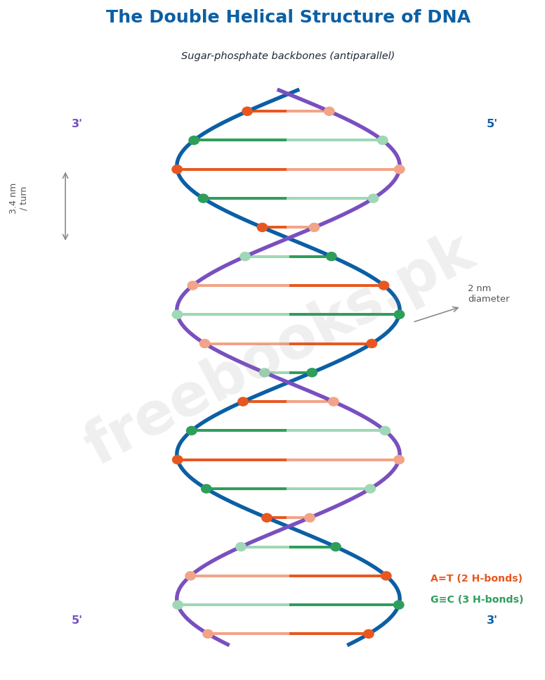
The significance of Chargaff's rule became clear when British chemist Rosalind Franklin, working in Maurice Wilkins's laboratory, produced an X-ray diffraction pattern of DNA fibres, revealing a helical structure with a diameter of 2 nm and a complete turn every 3.4 nm. Learning informally of Franklin's results before their 1953 publication, James Watson and Francis Crick at Cambridge University rapidly worked out the now-accepted DNA structure: a double helix of two antiparallel strands (one running 3' to 5', the other 5' to 3'), with bases pointing inward and pairing such that a large purine always pairs with a small pyrimidine, keeping the helix diameter constant at 2 nm; adenine forms two hydrogen bonds with thymine, and guanine forms three hydrogen bonds with cytosine (explaining Chargaff's equal A=T and G=C ratios), with base pairs stacked 0.34 nm apart, stabilised by hydrophobic interactions.
Semi-conservative DNA Replication and the Meselson-Stahl Experiment
The Watson-Crick model immediately suggested that DNA replication relies on complementarity: if the double helix is 'unzipped,' each exposed single strand can serve as a template for assembling a new complementary strand, producing two daughter duplexes each identical in sequence to the original. This mechanism, in which each daughter duplex contains one original (parental) strand and one newly synthesised strand, is called semi-conservative replication, since the original sequence is conserved but the original duplex itself is not (its two strands separate to form parts of two different new duplexes). Two rival hypotheses were also proposed: the conservative model, in which the original duplex would remain fully intact and generate an entirely separate new duplex, and the dispersive model, in which each strand of every daughter molecule would be a patchwork mixture of old and new DNA.
These three hypotheses were tested in 1958 by Matthew Meselson and Franklin Stahl. They grew bacteria in a medium containing heavy nitrogen (15N), labelling their DNA, then transferred the bacteria to a medium with normal, lighter nitrogen (14N) and collected DNA samples at intervals, separating them by density using cesium chloride density-gradient ultracentrifugation. DNA collected immediately after transfer was uniformly dense (all 15N); after one round of replication in 14N medium, all the DNA had an intermediate density; after a second round, two density classes appeared, one intermediate and one matching pure 14N DNA. This pattern was exactly as predicted by semi-conservative replication: after the first round, every daughter duplex is a hybrid of one heavy and one light strand, and when a hybrid duplex itself replicates, it produces one new hybrid duplex and one new fully light duplex, definitively confirming the Watson-Crick model's semi-conservative replication mechanism and ruling out the conservative and dispersive models.
The Replication Process: Enzymes and Mechanism
DNA replication begins at specific origin sequences on the DNA molecule, where DNA polymerase III and associated enzymes catalyse addition of nucleotides to growing complementary strands. Bacteria have three DNA polymerases (I, II and III); DNA polymerase I plays a small supporting role, while DNA polymerase III, a large dimeric enzyme, is the true replicating enzyme, threading DNA through itself at about 1000 nucleotides per second. DNA polymerase III can only extend an existing nucleotide chain already paired with the template, so it cannot initiate synthesis alone; instead, the enzyme primase first constructs a short RNA primer (about 10 nucleotides) complementary to the template, which DNA polymerase III then extends with DNA nucleotides, with the RNA primer nucleotides later replaced by DNA.
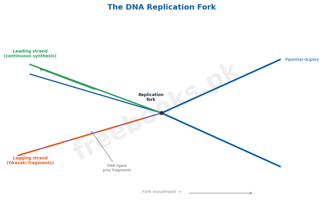
DNA polymerase III can only add nucleotides to the 3' end of a strand, so replication always proceeds in the 5' to 3' direction; because the two parental strands are antiparallel, the two new strands must be synthesised differently. The leading strand, elongating toward the replication fork, is synthesised continuously by simply adding nucleotides to its growing 3' end. The lagging strand, elongating away from the replication fork, is synthesised discontinuously as a series of short Okazaki fragments (about 100-200 nucleotides in eukaryotes, 1000-2000 in prokaryotes), each begun with its own RNA primer and synthesised 5' to 3' away from the fork; when DNA polymerase III reaches the previous fragment, DNA ligase joins the fragments together, while the enzyme jumps ahead to begin the next fragment as the DNA continues to unwind.
What Is a Gene? Garrod and the One Gene/One Polypeptide Hypothesis
In 1902, Archibald Garrod and William Bateson found certain diseases clustering in particular families, behaving as simple recessive Mendelian traits; investigating alkaptonuria, in which patients' urine darkens on exposure to air due to accumulated homogentisic acid, Garrod concluded that affected patients lacked the enzyme needed to break this substance down, and speculated that many inherited diseases might reflect similar enzyme deficiencies – implying that chromosomal DNA specifies particular enzymes, though this was not definitively established until 1941. Stanford geneticists George Beadle and Edward Tatum irradiated Neurospora (bread mould) spores with X-rays to induce mutations, then grew the progeny on a 'minimal' medium containing only basic nutrients; mutants unable to synthesise a required compound failed to survive unless that specific compound was added back, allowing precise identification of each mutant's biochemical deficiency. Adding arginine, for example, rescued several mutant strains ('arg mutants'), whose mutations clustered at three distinct chromosomal locations, one for each enzyme in the arginine biosynthetic pathway.
Since each enzyme defect traced to a mutation at a single, distinct chromosomal site, Beadle and Tatum concluded that genes act by specifying enzyme structure, with each gene encoding one enzyme – the one gene/one enzyme hypothesis, now more commonly called one gene/one polypeptide, since many enzymes consist of multiple polypeptide subunits each encoded by a separate gene. Since enzymes catalyse the assembly of all cellular components (nucleic acids, proteins, carbohydrates, lipids), DNA's control of enzyme structure ultimately specifies the structure of the organism itself. In 1953, Frederick Sanger sequenced the amino acids of insulin, proving proteins are definable, ordered chains of amino acids; in 1956, Vernon Ingram showed sickle cell anaemia results from a single amino acid change (glutamic acid to valine) in haemoglobin, caused by a single DNA base change (thymine to adenine) – establishing that a gene is the sequence of nucleotides that determines the amino acid sequence of a protein.
Central Dogma: Transcription and the Genetic Code
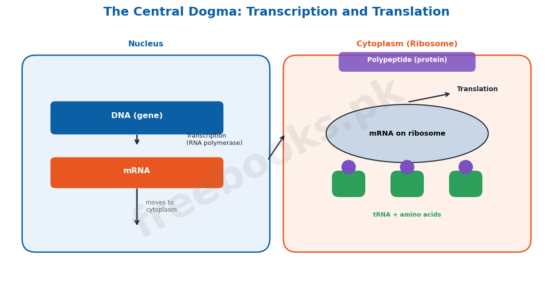
All organisms follow the same basic mechanism for reading and expressing genes, called the central dogma: genetic information flows from DNA to RNA (transcription) and then from RNA to protein (translation). Transcription begins when RNA polymerase binds a promoter sequence upstream of a gene, then moves along the DNA synthesising a complementary mRNA strand until it reaches a stop signal and releases the newly made RNA; only one DNA strand (the template or antisense strand) is transcribed, while the other (coding or sense strand) matches the mRNA sequence. Prokaryotes have a single RNA polymerase making all three RNA types, while eukaryotes have three: RNA polymerase I (rRNA), II (mRNA), and III (tRNA). In eukaryotes, the newly made mRNA is modified with a 7-methyl-GTP cap at the 5' end and a poly-A tail at the 3' end, protecting it during its journey from nucleus to ribosome and stabilising it against degradation.
There are three major classes of RNA: ribosomal RNA (rRNA), which forms part of the ribosome and provides the site of polypeptide assembly; transfer RNA (tRNA), which transports amino acids to the ribosome and positions them correctly on the growing polypeptide (humans have about 45 different tRNAs); and messenger RNA (mRNA), transcribed from DNA and carrying the code for protein synthesis to the ribosome. The genetic code consists of three-nucleotide codons, since a two-nucleotide code (4² = 16 combinations) cannot specify all 20 common amino acids, while a three-nucleotide code (4³ = 64 combinations) provides more than enough; the code was fully determined in the mid-1960s through experiments by Marshall Nirenberg, Philip Leder and Har Gobind Khorana. Of the 64 codons, three (UAA, UAG, UGA) are nonsense/stop codons that terminate translation, while AUG serves as the universal start codon (encoding methionine); the genetic code is nearly universal across all organisms (allowing genes to be transferred between species and still function), though minor exceptions exist in mitochondrial DNA, where, for example, UGA codes for tryptophan rather than acting as a stop codon.
Translation and Mutations
Translation begins when the initial portion of an mRNA binds to a ribosome, exposing one codon at a time at the ribosome's peptidyl (P) site; a tRNA molecule carrying the complementary anticodon and its specific attached amino acid binds each exposed codon in turn, with aminoacyl-tRNA synthetase enzymes (one for each of the 20 amino acids) ensuring each tRNA carries the correct amino acid. Translation begins with formation of an initiation complex, in which a special initiator tRNA carrying methionine binds the small ribosomal subunit at the P site, positioned by initiation factors, before the complex binds the mRNA's AUG start codon; the large ribosomal subunit then joins, and successive aminoacyl-tRNAs bind the exposed codon at the adjacent A (aminoacyl) site, with each new amino acid joined to the growing chain by a peptide bond as the ribosome translocates three nucleotides along the mRNA (5' to 3') for each cycle, shifting the growing chain to the P site and ejecting the spent tRNA at the E (exit) site; elongation continues until a nonsense (stop) codon is reached, which is recognised not by a tRNA but by release factor proteins that free the completed polypeptide from the ribosome.
If the DNA in all cells of an adult human were laid end to end, it would stretch nearly 100 billion kilometres, roughly 60 times the Earth-Jupiter distance, so even small changes (mutations), whether from replication errors or damage to the genetic message, can have significant effects; mutations in somatic cells are not inherited and have limited evolutionary consequence, while mutations in germ-line cells are passed to offspring, providing the raw material for evolutionary change through natural selection. Mutations are broadly classified as chromosomal aberrations, large-scale changes such as an extra or missing chromosome, or structural changes like deletions, insertions or inversions (causing syndromes such as Down's syndrome or Klinefelter's syndrome), and point mutations, small changes affecting only one or a few DNA base pairs, arising either from spontaneous replication errors or from damage by mutagens such as radiation or chemicals. Sickle cell anaemia (a single glutamic acid to valine substitution in haemoglobin) and phenylketonuria (a defective phenylalanine hydroxylase enzyme, causing toxic phenylalanine accumulation and mental retardation if untreated) are classic, well-studied examples of point mutations with serious clinical consequences.
Important Definitions
Karyotype
The particular array of chromosomes (number, size, shape) that characterizes an individual or species.
Transformation
The transfer of genetic material from one cell to another, altering the genetic makeup of the recipient cell (demonstrated by Griffith).
Semi-conservative replication
The mode of DNA replication in which each daughter duplex contains one original (parental) strand and one newly synthesized strand.
Okazaki fragment
A short DNA segment (100-200 nucleotides in eukaryotes) synthesized discontinuously on the lagging strand during DNA replication.
Gene
The sequence of nucleotides in DNA that determines the amino acid sequence of a protein (or specifies a functional RNA).
Transcription
The process by which an RNA copy of a gene's DNA sequence is synthesized by RNA polymerase.
Translation
The process by which the nucleotide sequence of mRNA is used by ribosomes to direct synthesis of a polypeptide's amino acid sequence.
Point mutation
A mutation involving a change in only one or a few base pairs in the DNA coding sequence.
Key Facts
| Item | Fact |
|---|---|
| Human chromosome number | Human cells have 46 chromosomes, in 23 homologous pairs. |
| DNA content per human chromosome | A typical human chromosome contains about 140 million (1.4 x 10⁸) nucleotides, about 5 cm long if stretched out. |
| Nucleosome spacing | The DNA duplex coils around a core of 8 histone proteins every 200 nucleotides, forming a nucleosome. |
| DNA helix dimensions | The DNA double helix has a diameter of 2 nm, with a complete turn every 3.4 nm; base pairs stack 0.34 nm apart. |
| Base pairing (hydrogen bonds) | Adenine pairs with thymine via 2 hydrogen bonds; guanine pairs with cytosine via 3 hydrogen bonds. |
| DNA polymerase III speed | DNA polymerase III adds nucleotides at a rate of about 1000 nucleotides per second. |
| Genetic code | The genetic code is a triplet (3-nucleotide) code; 4³ = 64 possible codons specify 20 amino acids, with 3 stop codons (UAA, UAG, UGA) and 1 start codon (AUG = methionine). |
| tRNA diversity | Human cells contain about 45 different kinds of tRNA molecules. |
Diagrams & Illustrations
The Double Helical Structure of DNA: a labelled diagram of the Watson-Crick DNA double helix, showing the antiparallel sugar-phosphate backbones, complementary base pairing (A=T with 2 hydrogen bonds, G≡C with 3 hydrogen bonds), and helix dimensions (2 nm diameter, 3.4 nm per turn).
The DNA Replication Fork: a labelled diagram of a DNA replication fork showing the leading strand (synthesized continuously toward the fork) and lagging strand (synthesized discontinuously as Okazaki fragments, joined by DNA ligase, away from the fork).
The Central Dogma: Transcription and Translation: a flow diagram showing the central dogma of gene expression: DNA is transcribed into mRNA (transcription) in the nucleus, and mRNA is translated into a polypeptide chain by ribosomes and tRNA (translation) in the cytoplasm.
Short Questions & Answers
What is a karyotype, and what features of chromosomes does it describe?
A karyotype is the particular array of chromosomes an individual or species possesses; it describes chromosome number, size, staining properties, centromere position, and relative arm lengths, and can differ markedly even among individuals of the same species.
Briefly describe Griffith's transformation experiment and its significance.
Griffith found that mixing dead virulent S bacteria with live harmless R bacteria killed mice, and live virulent S bacteria were recovered from them; he concluded genetic information had passed from the dead S bacteria to the living R bacteria (transformation), providing the first evidence that a hereditary 'transforming principle' exists, later identified as DNA by Avery.
How did the Hershey-Chase experiment confirm DNA (not protein) is the hereditary material?
By labelling viral DNA with radioactive 32P and viral protein coats with radioactive 35S, Hershey and Chase showed that only the 32P (DNA) label entered infected bacteria and appeared in the next generation of viruses, while the 35S (protein) label stayed outside, proving DNA carries the hereditary information.
State Chargaff's rule and explain how it relates to the Watson-Crick base pairing model.
Chargaff's rule states that in DNA, the amount of adenine always equals thymine, and guanine always equals cytosine; this is explained by Watson-Crick base pairing, in which adenine always pairs specifically with thymine (2 H-bonds) and guanine always pairs specifically with cytosine (3 H-bonds), so the two bases in each pair must always occur in equal amounts.
Differentiate the leading strand and lagging strand during DNA replication.
The leading strand elongates continuously toward the replication fork, with nucleotides added directly to its growing 3' end; the lagging strand elongates away from the fork and is synthesized discontinuously as short Okazaki fragments, each requiring its own RNA primer, later joined together by DNA ligase.
Differentiate a chromosomal aberration and a point mutation, with one example of each.
A chromosomal aberration is a large-scale change, such as an extra or missing chromosome or a deletion/insertion/inversion of a chromosome segment (e.g. Down's syndrome); a point mutation is a small change affecting only one or a few DNA base pairs (e.g. sickle cell anaemia, caused by a single base change altering one amino acid in haemoglobin).
Long Questions & Answers
Describe the Meselson-Stahl experiment and explain how it demonstrated that DNA replicates semi-conservatively.
The Watson-Crick model of DNA structure immediately suggested a natural mechanism for copying genetic information: if the two strands of the double helix were separated, each could serve as a template along which a new complementary strand could be assembled, producing two daughter duplexes each identical in sequence to the original parent molecule; this proposed mechanism was called semi-conservative replication, because while the original nucleotide sequence would be faithfully conserved across generations, the original physical duplex itself would not remain intact, since each of its two strands would end up as part of a different new duplex. However, semi-conservative replication was not the only hypothesis proposed to explain how DNA might be copied: the conservative model held that the entire original parental double helix would remain completely intact after replication, with an entirely new, separate daughter duplex generated alongside it, while the dispersive model predicted an even more thorough mixing, in which the parental DNA would become fully dispersed throughout all the daughter molecules, so that every single strand in every daughter duplex would actually be a patchwork mixture of old, original DNA and newly synthesized DNA. To distinguish decisively between these three competing hypotheses, Matthew Meselson and Franklin Stahl, working at the California Institute of Technology, designed an elegant experiment in 1958 using different isotopes of nitrogen as density labels. They first grew several generations of bacteria in a growth medium containing only the heavy isotope of nitrogen, 15N, which became incorporated throughout the nitrogen-containing bases of all the bacterial DNA, making that DNA measurably denser than normal DNA; once the bacterial DNA was uniformly heavy, they abruptly transferred the bacterial culture into a fresh medium containing only the ordinary, lighter isotope of nitrogen, 14N, and then carefully collected samples of the bacterial DNA at successive time intervals as the bacteria continued to grow and divide, replicating their DNA using the now-available lighter nitrogen. To measure the density of the DNA collected at each interval, they dissolved each sample in a concentrated cesium chloride solution and spun it at extremely high speed in an ultracentrifuge; the intense centrifugal force caused the heavy cesium ions themselves to migrate toward the bottom of the tube, establishing a stable density gradient within the tube, and any given DNA molecule would migrate through this gradient and come to rest at exactly the point where its own density matched the surrounding cesium chloride solution, with denser 15N-containing DNA settling further down the tube than lighter 14N-containing DNA. The results were striking and unambiguous: the DNA sample collected immediately at the moment of transfer into the 14N medium was uniformly dense, forming a single band at the position expected for pure 15N DNA, exactly as expected since no replication had yet occurred in the new medium. However, after the bacteria had completed exactly one full round of DNA replication in the 14N medium, all of the resulting DNA now formed a single band at an intermediate density, exactly halfway between the density expected for pure 15N DNA and pure 14N DNA, a result immediately inconsistent with the conservative model (which would have predicted two separate bands, one still fully heavy and one fully light, rather than a single uniform intermediate band). Then, after the bacteria completed a second full round of replication, two distinct density bands appeared simultaneously: one band still at the intermediate density observed after the first round, and a second, new band at the density of pure, fully light 14N DNA, a result that decisively ruled out the dispersive model as well, since dispersive replication would have predicted that all the DNA converge toward a single density approaching pure 14N DNA rather than splitting cleanly into two distinct density classes. Meselson and Stahl correctly interpreted this precise pattern as the direct signature of semi-conservative replication: after the first round of replication, every single daughter DNA duplex was necessarily a hybrid molecule, containing exactly one original heavy 15N strand paired with one newly synthesized light 14N strand, which is why all the DNA appeared at a single, uniform intermediate density at that stage; then, when each of these hybrid duplexes itself underwent a second round of replication, each hybrid duplex separated into its two individual strands, with the original heavy 15N strand serving as a template to synthesize a new light partner strand (regenerating another hybrid, intermediate-density duplex), while the light 14N strand from the first round separately served as a template to synthesize its own new light partner strand (producing an entirely new, fully light duplex) – together producing exactly the two density bands actually observed, providing clear, direct experimental confirmation of the semi-conservative replication mechanism originally predicted by the Watson-Crick model.
Describe the process of transcription in detail, and explain how the genetic code specifies the sequence of amino acids in a protein.
Transcription is the first step of the central dogma of gene expression, the process by which the genetic information encoded in a gene's DNA sequence is copied into a complementary strand of messenger RNA (mRNA), and it proceeds through a precise, well-regulated sequence of molecular events. Only one of the two DNA strands making up a gene actually serves as the template for RNA synthesis at any given time, and this strand is specifically called the template strand or antisense strand, while the opposite, non-transcribed DNA strand, which carries the same sequence as the resulting mRNA (except for using thymine in place of uracil), is called the coding strand or sense strand. The process begins when the enzyme RNA polymerase specifically recognizes and binds to a particular DNA sequence located just upstream of the gene itself, called the promoter; in prokaryotes, this promoter region contains two short, specific binding sequences that have a particular affinity for RNA polymerase, one located around position -35 (with the consensus sequence TTGACA) and another around position -10 (with the consensus sequence TATAAT), while in eukaryotic promoters, functionally analogous binding sites are instead found further upstream, at approximately positions -75 and -25. Once RNA polymerase has bound correctly to the promoter, guided in bacteria by a specialized subunit called the sigma factor (which is responsible specifically for ensuring correct initiation of transcription and is subsequently released once transcription is properly underway), the remaining core enzyme complex proceeds to move steadily along the template strand of the gene; as it advances, the two strands of the DNA double helix are locally and transiently separated or 'unzipped' at the exact point where the enzyme is actively working, creating a small, characteristic bubble-like structure called the transcription bubble, which itself progressively moves down the length of the gene as transcription proceeds, with the newly synthesized RNA strand visibly protruding from the moving bubble as it lengthens. RNA polymerase continues synthesizing RNA in a fixed 5' to 3' direction, continuously adding nucleotides that are complementary to the template DNA strand, until it eventually reaches a specific stop or termination sequence positioned at the far end of the gene; in many cases, this termination sequence consists of a stretch of GC-rich base pairs immediately followed by a stretch of AT-rich base pairs, and the RNA transcript itself, once synthesized from this particular region, is able to fold back on itself to form a stable hairpin loop structure, followed immediately by a run of four or more uracil ribonucleotides, and this specific combination of a hairpin structure followed by U residues causes RNA polymerase to physically stall, disengage completely from the DNA template, and release the finished RNA transcript. Once this newly transcribed mRNA molecule has been produced, its genetic information is subsequently decoded and converted into a specific sequence of amino acids according to the genetic code, a universal biological cipher in which each successive, non-overlapping group of exactly three nucleotides, called a codon, specifies one particular amino acid to be incorporated into the growing protein chain; three nucleotides are required for this coding system specifically because a code based on only two nucleotides at a time could generate at most 4 squared, or 16, distinct combinations, which is insufficient to uniquely specify all 20 amino acids commonly found in proteins, whereas a three-nucleotide code generates 4 cubed, or a full 64 distinct possible combinations, more than enough to comfortably cover all 20 amino acids with some redundancy remaining. Of these 64 possible codons, 61 each specify one of the 20 standard amino acids (with most amino acids therefore specified by more than one alternative codon), while the remaining three codons, UAA, UAG and UGA, do not correspond to any amino acid at all and instead function specifically as stop or nonsense codons that signal the definitive end of the coding sequence and terminate protein synthesis; in addition, one particular codon, AUG, serves the important dual role of both specifying the amino acid methionine and simultaneously acting as the universal start codon that marks precisely where translation of every protein-coding sequence must begin. Because this fundamental three-nucleotide codon system, and the specific assignment of particular codons to particular amino acids, is very nearly identical and universally shared across almost all known organisms, from simple bacteria through to complex eukaryotes such as humans, it becomes possible in principle to experimentally transfer a functioning gene from one organism into an entirely different host organism and still have that transferred gene correctly transcribed and translated into the very same intended protein by the new host's own cellular machinery, a fact of tremendous practical importance underlying essentially the entire modern field of genetic engineering and biotechnology.
MCQs with Answers
Chromosomes were first observed by Walther Flemming while examining dividing cells of: (a) fruit flies (b) salamander larvae (c) bacteria (d) pea plants
Correct Answer: (b) salamander larvae. Flemming first observed chromosomes in 1882 while examining rapidly dividing cells of salamander larvae.
The chromosomal theory of inheritance was first formulated in 1902 by: (a) Karl Correns (b) Thomas Hunt Morgan (c) Walter Sutton (d) Gregor Mendel
Correct Answer: (c) Walter Sutton. Walter Sutton formulated the chromosomal theory of inheritance in 1902, based on parallels between chromosome behavior and Mendelian inheritance.
Griffith's experiment demonstrating 'transformation' used which bacterium? (a) Escherichia coli (b) Streptococcus pneumoniae (c) Neurospora crassa (d) Salmonella typhi
Correct Answer: (b) Streptococcus pneumoniae. Griffith worked with Streptococcus pneumoniae (then Pneumococcus), using virulent (S) and non-virulent (R) strains.
The Hershey-Chase experiment used radioactive isotopes 32P and 35S to label, respectively: (a) protein and DNA (b) DNA and protein (c) RNA and DNA (d) protein and RNA
Correct Answer: (b) DNA and protein. 32P labelled phage DNA (phosphate-containing) and 35S labelled phage protein coats (sulfur-containing amino acids).
According to Chargaff's rule, in any DNA sample the amount of adenine always equals the amount of: (a) guanine (b) cytosine (c) thymine (d) uracil
Correct Answer: (c) thymine. Chargaff's rule states A always equals T, and G always equals C, in double-stranded DNA.
In the DNA double helix, guanine pairs with cytosine via how many hydrogen bonds? (a) one (b) two (c) three (d) four
Correct Answer: (c) three. Guanine pairs with cytosine via three hydrogen bonds, while adenine pairs with thymine via two.
The Meselson-Stahl experiment confirmed that DNA replication is: (a) conservative (b) dispersive (c) semi-conservative (d) non-replicative
Correct Answer: (c) semi-conservative. Meselson and Stahl's density-gradient results matched the predictions of semi-conservative replication, ruling out conservative and dispersive models.
On the lagging strand, DNA is synthesized discontinuously as short segments called: (a) primers (b) Okazaki fragments (c) nucleosomes (d) codons
Correct Answer: (b) Okazaki fragments. The lagging strand is synthesized as short Okazaki fragments, later joined together by DNA ligase.
Beadle and Tatum's experiments on Neurospora led to which hypothesis? (a) one gene/one chromosome (b) one gene/one polypeptide (c) one gene/one codon (d) one codon/one nucleotide
Correct Answer: (b) one gene/one polypeptide. Beadle and Tatum's arginine mutant studies established the one gene/one enzyme (now one gene/one polypeptide) hypothesis.
Which of the following is a stop (nonsense) codon in the genetic code? (a) AUG (b) UGG (c) UAA (d) AUA
Correct Answer: (c) UAA. UAA is one of three stop/nonsense codons (along with UAG and UGA) that terminate translation; AUG is the start codon.
Quick Revision Summary
- Chromosome basics: humans = 46 (23 pairs). Structure: chromatid + centromere + secondary constriction. Types by centromere position: telocentric/acrocentric/submetacentric/metacentric. Composition: ~40% DNA + 60% protein. Nucleosome = DNA + 8 histones (every 200 nucleotides). Heterochromatin (permanently condensed) vs Euchromatin (condenses only during division).
- Chromosomal theory: Sutton (1902, chromosome-gene parallel) -> Morgan (1910, Drosophila white-eye = sex-linked, gene on X chromosome) = proved genes are ON chromosomes.
- DNA = hereditary material: Griffith (1928, transformation, dead S + live R -> live S) -> Avery/MacLeod/McCarty (1944, DNase destroys transforming activity = DNA is the agent) -> Hershey-Chase (1952, 32P-DNA enters bacteria, 35S-protein stays outside) = confirmed DNA.
- DNA structure: Miescher (1869, discovered 'nuclein') -> Levene (1920s, nucleotide = sugar+phosphate+base) -> Chargaff (A=T, G=C) -> Franklin (X-ray diffraction, helix, 2nm diameter/3.4nm turn) -> Watson & Crick (1953, double helix, antiparallel strands, A=T 2 H-bonds, G≡C 3 H-bonds).
- Replication: Semi-conservative (Meselson-Stahl 1958, 15N/14N density gradient, confirmed hybrid bands). Process: origin -> primase makes RNA primer -> DNA polymerase III extends 5'->3' (1000 nt/sec) -> Leading strand (continuous) vs Lagging strand (Okazaki fragments + DNA ligase).
- Gene concept: Garrod (1902, alkaptonuria = enzyme deficiency) -> Beadle & Tatum (1941, Neurospora, arg mutants) = one gene/one polypeptide. Sanger (1953, insulin sequence) + Ingram (1956, sickle cell = Glu->Val mutation) = gene defines protein sequence.
- Central dogma: DNA -> (transcription, RNA polymerase, promoter) -> mRNA -> (translation, ribosome+tRNA) -> protein. 3 RNA types: mRNA/tRNA/rRNA. Genetic code: triplet, 64 codons, AUG=start, UAA/UAG/UGA=stop, nearly universal. Mutations: Chromosomal aberration (large-scale, e.g. Down's) vs Point mutation (1-few bases, e.g. sickle cell/PKU). Notes by freebooks.pk.
Exam Tips
- Memorise the three DNA-is-the-hereditary-material experiments as a linked chain with one key result each: Griffith (transformation happens) -> Avery (DNase destroys it = it's DNA) -> Hershey-Chase (32P/DNA enters cells, 35S/protein doesn't).
- Keep Chargaff's rule and Watson-Crick base pairing as one linked fact: A=T (2 H-bonds), G=C (3 H-bonds) – this single fact answers many 'why' questions about DNA structure and replication.
- Draw and label the semi-conservative replication diagram from memory (parent duplex -> two hybrid duplexes -> one hybrid + one fully new duplex) – this is one of the most commonly asked long questions.
- Build a 2-column comparison table for leading vs lagging strand (continuous vs discontinuous, toward vs away from fork, no fragments vs Okazaki fragments) rather than memorising as separate facts.
- Learn the one gene/one polypeptide chain of evidence in order: Garrod (enzyme deficiency idea) -> Beadle & Tatum (Neurospora proof) -> Sanger (protein = amino acid sequence) -> Ingram (sickle cell = 1 amino acid change).
- Memorise the central dogma as a simple flow: DNA –transcription–> mRNA –translation–> Protein, and know which RNA polymerase makes which RNA in eukaryotes (I=rRNA, II=mRNA, III=tRNA).
- For the genetic code, remember the '4-16-64' logic (4 bases, 2-letter code=16 combos insufficient, 3-letter code=64 combos sufficient) rather than just memorising 'triplet code' as an isolated fact – this explains 'why triplet' questions.