This chapter covers The Cell from the 1st Year (FSc Part-I) Biology syllabus of the Punjab Curriculum and Textbook Board (PTB/PCTB). The cell is the structural and functional unit of life — the smallest unit that can carry out all the activities of a living organism. The chapter traces how the Cell Theory emerged from Robert Hooke's first observations of cork in 1665 through Schwann, Schleiden and Virchow, and then works through the structure of a generalized eukaryotic cell: the plasma membrane, cytoplasm, and every major organelle. These notes are prepared by freebooks.pk.
It closes by comparing prokaryotic and eukaryotic cells, the most fundamental division in all of biology. Understanding the parts of a cell and how they work together is essential background for cell division, genetics, and every later chapter that deals with how organisms grow, reproduce and carry out their metabolism.
Learning Objectives
- State the Cell Theory and describe its historical development.
- Describe the structure and function of the plasma membrane, including the Fluid Mosaic Model.
- Describe the structure and function of the endoplasmic reticulum, ribosomes, Golgi apparatus and lysosomes.
- Describe peroxisomes, glyoxysomes, vacuoles, the cytoskeleton and centrioles.
- Describe the structure of mitochondria and explain why they are called the powerhouse of the cell.
- Describe the three types of plastids and the structure of the chloroplast.
- Describe the structure of the nucleus, including the nuclear membrane, nucleolus and chromosomes.
- Differentiate between prokaryotic and eukaryotic cells.
Key Concepts
Cell Theory: History and Modern Statement
The cell can be defined as the structural and functional unit of life — the smallest unit that can carry out all the activities of life, and the building block of complex multicellular organisms. Cell biology began with Robert Hooke (1665), who examined thin sections of cork under his own compound microscope and, in his book Micrographia, described the honeycomb-like compartments he called cells, though he thought of a cell as simply an empty space bounded by walls. In 1831 Robert Brown reported the presence of the nucleus, showing a cell was not empty, and in 1838-39 the botanist Schleiden and the zoologist Schwann, working independently, proposed the Cell Theory: that cells consist of a nucleus, cytoplasm and an outer plasma membrane (with a cell wall as an additional plant-only structure), and that all living organisms are composed of cells and cell products.
The theory was later extended by Rudolph Virchow (1855), who proposed that new cells arise only by the division of pre-existing cells ("omnis cellula e cellula"), a view supported experimentally by Louis Pasteur (1862), who showed bacteria arise only from existing bacteria, disproving spontaneous generation. August Weismann (1880) added that all living cells share a common origin because of their basic similarities in structure and molecules. The modern Cell Theory states: all organisms are composed of one or more cells; all cells arise from pre-existing cells; and the cell is the basic structural and functional unit of all organisms. The development of microscopy has driven this understanding further — the naked eye can resolve points about 1.0 mm apart, a compound microscope improves this to about 2.0 micrometres (500 times better), and an electron microscope reaches a resolution of 2-4 Angstrom units, about 250,000 times better than the naked eye, revealing the fine internal structure of cell organelles.
The Plasma Membrane and Cell Wall
The plasma membrane (cell membrane) is the outermost boundary of the cell, chemically composed of about 60-80% protein and 20-40% lipid, plus a small amount of carbohydrate. Earlier models proposed a lipid bilayer sandwiched between two continuous protein layers (the unit membrane), but later evidence showed the protein is not continuous but embedded within the lipid layer in a mosaic pattern, leading to the now widely accepted Fluid Mosaic Model. The membrane is differentially (selectively) permeable, letting lipid-soluble and small neutral molecules such as gases, water and glucose cross more easily than charged ions; movement down a concentration gradient is passive, while movement against a gradient requires energy from ATP and is called active transport. Some animal cells also take in materials by endocytosis, which may be phagocytosis (engulfing solid particles) or pinocytosis (taking in liquid).
In most plant cells, an additional outer boundary, the cell wall, lies outside the plasma membrane, secreted by the cell's protoplasm and composed of a primary wall (cellulose with some pectin and hemicellulose), a secondary wall (thicker, rigid, with inorganic salts, silica, waxes, cutin and lignin), and the middle lamella between neighbouring cells. Unlike the plasma membrane, the cell wall does not block materials from passing through; its main role is to give the cell a definite, rigid shape. A bacterial cell wall is chemically different, built of peptidoglycan (murein) rather than cellulose, while fungal cell walls are made of chitin.
Cytoplasm and the Endomembrane System
The living contents of a eukaryotic cell are divided into the nucleus and the cytoplasm, which together form the protoplasm. The soluble part of the cytoplasm, the cytosol, is about 90% water and forms the ground substance in which cell organelles are suspended; it also serves as a storehouse of vital chemicals and a site for processes such as glycolysis. The endoplasmic reticulum (ER) is a network of membrane channels running through the cytoplasm, continuous in places with both the plasma membrane and the nuclear membrane; the rough ER (RER) is studded with ribosomes and synthesizes proteins for storage or export, while the smooth ER (SER) lacks ribosomes and instead handles lipid metabolism, detoxifies harmful drugs, and in muscle and nerve cells helps transmit impulses.
Ribosomes are tiny ribonucleoprotein granules, first studied by Palade (1955), made of roughly equal amounts of RNA and protein; a eukaryotic ribosome has a large 60S subunit and a small 40S subunit that join to form an 80S particle, and a group of ribosomes attached to one mRNA strand is called a polysome. New ribosomes are assembled in the nucleolus and exported to the cytoplasm. The Golgi apparatus, discovered by Golgi in 1898, is a stack of flattened membrane sacs (cisternae) that receives material budded from the SER at its forming face and releases finished, packaged secretory vesicles at its maturing face, modifying proteins and lipids by adding carbohydrate to form glycoproteins and glycolipids. Lysosomes, first isolated by De Duve (1949), are single-membrane sacs rich in hydrolytic enzymes that digest engulfed foreign material (phagocytosis) and worn-out cell parts (autophagy); several inherited storage diseases, such as Tay-Sachs disease, result from a missing lysosomal enzyme.
Other Cytoplasmic Organelles
Peroxisomes, isolated by De Duve and coworkers (1965), are single-membrane organelles about 0.5 micrometres across that contain hydrogen-peroxide-producing oxidases and catalase, found in both animal and plant cells. Glyoxysomes are similar plant-only organelles, abundant in germinating, lipid-rich seeds such as castor bean and soybean, where they run the glyoxylate cycle to convert stored fatty acids into carbohydrate for the growing seedling. Vacuoles occur in both animal and plant cells but are especially large in plant cells, where a single central vacuole can occupy most of the cell volume; bounded by a single membrane, they store water and cell products and generate the turgor pressure that supports plant tissues.
The cytosol also contains a cytoskeleton built of three fibre types: microtubules (long tubulin structures involved in spindle formation during mitosis, and in cilia, flagella and centrioles), microfilaments (slender actin structures responsible for cyclosis and amoeboid movement), and intermediate filaments (which maintain cell shape). Centrioles, found in animal cells and some lower plants, are paired cylindrical structures near the nucleus, each made of nine microtubule triplets arranged at right angles to its partner; they duplicate before cell division and help organize the spindle and, in some cells, the formation of cilia. Centrioles are absent in higher plants.
Mitochondria: The Powerhouse of the Cell
Mitochondria are bound by two membranes: a smooth outer membrane and an inner membrane that folds inward as cristae, projecting into the central mitochondrial matrix; small knob-like F1 particles stud the inner surface of the cristae. The matrix contains enzymes, coenzymes and salts that carry out key metabolic pathways such as the Krebs cycle, aerobic respiration and fatty acid metabolism, converting the chemical energy of food into ATP, which the cell then uses on demand — this is why mitochondria are called the powerhouse of the cell. Mitochondria also contain their own DNA and ribosomes, meaning they can synthesize some of their own proteins and are self-replicating organelles; their number and size in a cell depend on how metabolically active that cell is.
Plastids: Chloroplasts, Chromoplasts and Leucoplasts
Plastids are membrane-bound, usually pigment-containing bodies found only in plant cells, of three main types. Chloroplasts, about 4-6 micrometres across, contain the green pigment chlorophyll (chemically similar to the haem of haemoglobin, but with magnesium instead of iron at its centre), which absorbs light energy for photosynthesis; under the electron microscope a chloroplast shows a double-membrane envelope, a fluid stroma (containing enzymes, ribosomes and a small circular DNA, where carbon dioxide is fixed into sugars), and stacks of flattened thylakoid membranes called grana (usually 50 or more thylakoids per granum), which is where light energy is trapped and ATP is generated; chloroplasts, like mitochondria, are self-replicating. Chromoplasts give flowers and ripe fruit their non-green colours, helping with pollination and seed dispersal, while leucoplasts are colourless plastids found in underground plant parts, where they store food.
The Nucleus: Membrane, Nucleolus and Chromosomes
The nucleus, first reported by Robert Brown in 1831, controls the life and activities of the cell; it is usually central in animal cells but pushed to the periphery in plant cells by the large vacuole. It is surrounded by a nuclear envelope made of two membranes, continuous with the ER at points, with nuclear pores where the two membranes meet, allowing regulated exchange of material between nucleus and cytoplasm (undifferentiated cells such as eggs may have about 30,000 pores per nucleus, while differentiated cells such as red blood cells have only 3 or 4). Inside, the nucleolus is a densely stained, non-membrane-bound body where ribosomal RNA is synthesized and ribosomal subunits are assembled before export through the nuclear pores.
In a non-dividing cell, hereditary material exists as a diffuse chromatin network within the nucleoplasm; during cell division, this chromatin condenses into visible, darkly staining chromosomes, each initially made of two identical chromatids joined at a centromere (the point of attachment for spindle fibres during division). A chromosome is built of DNA and protein and carries genes, the units that control cell activities and pass hereditary information to the next generation. The chromosome number is constant within a species: human body cells are diploid with 46 chromosomes, while human sperm and egg cells are haploid with 23; other examples include 8 in the fruit fly Drosophila, 16 in onion, 48 in potato and 14 in the garden pea.
Prokaryotic and Eukaryotic Cells
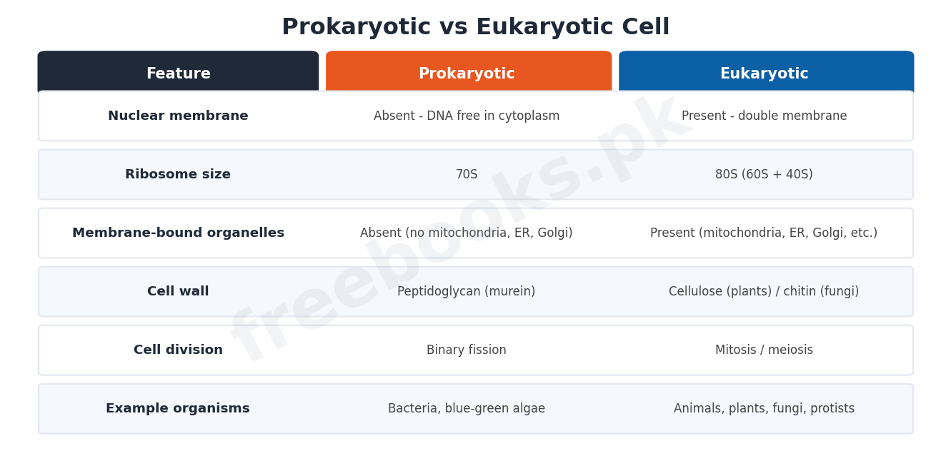
Cells are divided into two fundamental types based mainly on the structure of their nucleus. Eukaryotic cells (found in animals, plants, fungi and protists) have a well-defined nucleus in which the DNA is enclosed by a double nuclear membrane, along with membrane-bound organelles such as mitochondria, endoplasmic reticulum, Golgi apparatus and (in plants) chloroplasts. Prokaryotic cells (bacteria and blue-green algae) lack a nuclear membrane, so their DNA lies directly in the cytoplasm, and they also lack most of these membrane-bound organelles; prokaryotic ribosomes are smaller (70S) than eukaryotic ribosomes (80S), and since prokaryotes lack mitosis, they instead divide by binary fission. The most distinctive feature of the prokaryotic cell is its cell wall, built of peptidoglycan (murein) rather than the cellulose found in plant cell walls, and it was long thought (due to their simpler structure) that prokaryotic cells represent an earlier evolutionary stage than eukaryotic cells.
Important Definitions
Cell
The structural and functional unit of life; the smallest unit that can carry out all the activities of a living organism.
Fluid Mosaic Model
The currently accepted model of the plasma membrane, in which proteins are embedded in a mosaic pattern within a fluid lipid bilayer, rather than forming continuous outer layers.
Cytosol
The soluble, water-based part of the cytoplasm (about 90% water) in which cell organelles are suspended.
Polysome
A group of ribosomes attached to and translating the same strand of messenger RNA.
Mitochondrion
A double-membraned organelle that generates ATP through aerobic respiration; called the powerhouse of the cell.
Chloroplast
A plant-cell plastid containing chlorophyll, in which photosynthesis takes place.
Chromosome
A thread-like structure of DNA and protein, visible during cell division, that carries an organism's genes.
Prokaryotic cell
A cell, such as a bacterium, that lacks a nuclear membrane, so its DNA lies directly in the cytoplasm.
Key Facts
| Item | Fact |
|---|---|
| Cell discovery | Robert Hooke, 1665 – observed cork cells, published in Micrographia. |
| Cell Theory proposed | Schleiden and Schwann, 1838-39 (independently). |
| "Omnis cellula e cellula" | Rudolph Virchow, 1855 – new cells arise only from pre-existing cells. |
| Microscope resolution | Naked eye: ~1.0 mm. Compound microscope: ~2.0 micrometres. Electron microscope: 2-4 Angstrom. |
| Ribosome sedimentation | Eukaryotic: 80S (60S + 40S subunits). Prokaryotic: 70S. |
| Human chromosome number | Body cells (diploid): 46. Sperm/egg cells (haploid): 23. |
| Golgi apparatus discovery | Camillo Golgi, 1898. |
| Lysosome / Peroxisome discovery | De Duve: lysosomes (1949), peroxisomes (1965, with coworkers). |
Diagrams & Illustrations
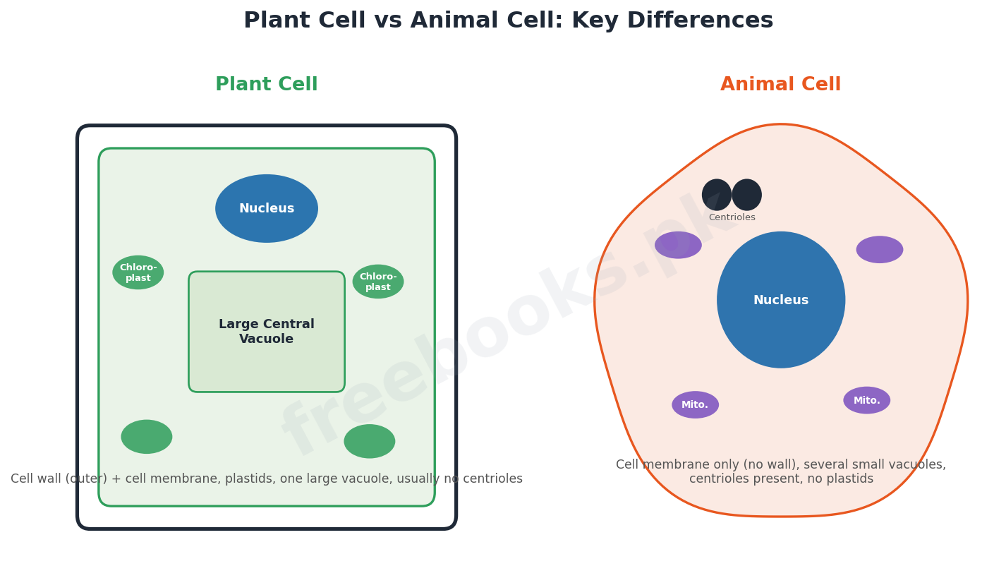
Plant Cell vs Animal Cell: Key Differences: a side-by-side comparison chart of a generalized plant cell and animal cell, highlighting structures present only in plant cells (cell wall, large central vacuole, plastids) versus structures typical of animal cells (centrioles, small scattered vacuoles).
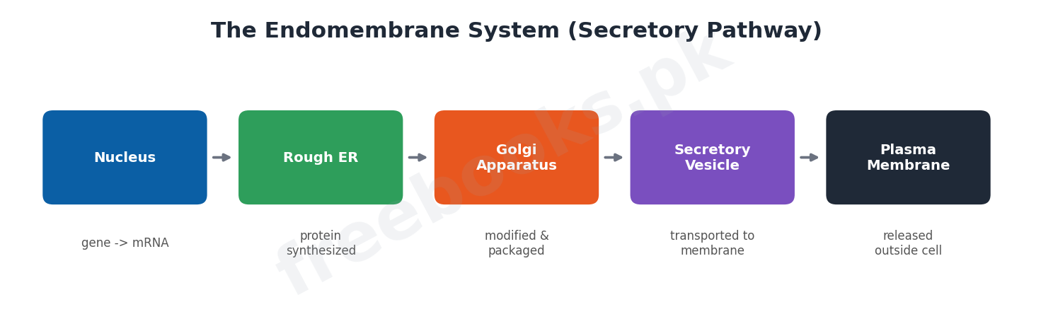
The Endomembrane System (Secretory Pathway): a flow diagram showing how a protein moves through the endomembrane system: synthesized on the rough endoplasmic reticulum, packaged and modified in the Golgi apparatus, then released as a secretory vesicle or stored in a lysosome.
Prokaryotic vs Eukaryotic Cell: a comparison chart of prokaryotic and eukaryotic cells across key features: nuclear membrane, ribosome size (70S vs 80S), membrane-bound organelles, and cell wall composition.
Short Questions & Answers
State the modern Cell Theory in three points.
All organisms are composed of one or more cells; all cells arise from pre-existing cells; and the cell is the basic structural and functional unit of all organisms.
Differentiate rough and smooth endoplasmic reticulum.
Rough endoplasmic reticulum (RER) has attached ribosomes and synthesizes proteins, while smooth endoplasmic reticulum (SER) lacks ribosomes and instead handles lipid metabolism, detoxification and impulse transmission in some cells.
What is the function of lysosomes? Name their discoverer.
Lysosomes digest engulfed foreign material and worn-out cell parts using hydrolytic enzymes; they were first isolated by De Duve in 1949.
Differentiate microtubules, microfilaments and intermediate filaments.
Microtubules are tubulin structures involved in spindle formation and organelles like cilia and centrioles; microfilaments are actin structures responsible for cell movement; intermediate filaments maintain cell shape.
State two differences between prokaryotic and eukaryotic cells.
Eukaryotic cells have a membrane-bound nucleus and 80S ribosomes, while prokaryotic cells have no nuclear membrane (DNA lies free in the cytoplasm) and smaller 70S ribosomes.
Why are mitochondria called the powerhouse of the cell?
Mitochondria carry out the Krebs cycle and aerobic respiration in their matrix, converting the chemical energy of food into ATP, which supplies energy for all cellular activities.
Long Questions & Answers
Describe the Cell Theory and discuss its historical emergence.
The Cell Theory developed gradually over two centuries. Robert Hooke first observed and named cells in 1665 while examining cork under a microscope, though he believed a cell was simply an empty space bounded by walls. Robert Brown's discovery of the nucleus in 1831 showed cells were not empty, and in 1838-39 Schleiden and Schwann, working independently, proposed that cells consist of a nucleus, cytoplasm and outer membrane, with a cell wall as an additional plant structure, and that all living organisms are made of cells and cell products. Rudolph Virchow extended the theory in 1855 with the idea that new cells arise only from pre-existing cells ("omnis cellula e cellula"), directly opposing the idea of spontaneous generation; Louis Pasteur supplied experimental proof of this in 1862 by showing bacteria arise only from existing bacteria. August Weismann later added that all living cells share a common origin, given their fundamental similarities in structure and chemistry. Together these contributions produced the modern Cell Theory: all organisms are composed of one or more cells, all cells arise from pre-existing cells, and the cell is the basic structural and functional unit of every organism — one of the most important unifying ideas in all of biology.
Describe the structure and function of the plasma membrane, including the Fluid Mosaic Model.
The plasma membrane is the outermost boundary of the cell (covered by a cell wall in most plant cells), chemically made of roughly 60-80% protein, 20-40% lipid, and a small amount of carbohydrate. Early models proposed that the membrane was a lipid bilayer sandwiched between two continuous protein layers, called the unit membrane; however, later evidence showed the protein is not continuous but embedded within the lipid bilayer in a scattered, mosaic arrangement, leading to the currently accepted Fluid Mosaic Model. Functionally, the plasma membrane is differentially (selectively) permeable: it acts as a barrier between the cell and its environment, allowing lipid-soluble molecules, gases, water and glucose to cross relatively freely, while restricting charged ions. Movement down a concentration gradient occurs passively, while movement against a gradient (from low to high concentration) requires energy supplied by ATP and is called active transport. In some animal cells, the membrane also folds inward to take in material by endocytosis — phagocytosis for solid particles or pinocytosis for liquids — and in neurons it transmits nerve impulses along the cell.
Compare the structure and function of mitochondria and chloroplasts.
Both mitochondria and chloroplasts are double-membrane-bound, self-replicating organelles that contain their own DNA and ribosomes, allowing them to synthesize some of their own proteins — a similarity that supports the idea that both organelles originated from ancient free-living bacteria. Structurally, a mitochondrion has a smooth outer membrane and a folded inner membrane (cristae) projecting into the central matrix, with small F1 particles on the cristae surface; the matrix carries out the Krebs cycle, aerobic respiration and fatty acid metabolism, converting food energy into ATP, which is why mitochondria are called the cell's powerhouse. A chloroplast, found only in plant cells, has a double-membrane envelope surrounding a fluid stroma (containing enzymes, ribosomes, and its own small circular DNA, where carbon dioxide is fixed into sugar) and stacks of thylakoid membranes called grana, which hold the green pigment chlorophyll and are the site where light energy is trapped and used to generate ATP during photosynthesis. In short, mitochondria release the chemical energy stored in food as usable ATP in almost all eukaryotic cells, while chloroplasts capture light energy and convert it into chemical energy stored in sugars, but only in photosynthetic plant cells.
Describe the structure of the nucleus, including the nuclear membrane, nucleolus and chromosomes.
The nucleus, discovered by Robert Brown in 1831, is usually the most prominent structure in a cell and controls all of its life and activities; it sits centrally in most animal cells but is pushed to one side in plant cells by the large central vacuole. It is enclosed by a nuclear envelope made of two membranes, which is continuous with the endoplasmic reticulum at certain points and perforated by nuclear pores where the outer and inner membranes meet, allowing regulated exchange of material with the cytoplasm; undifferentiated cells such as eggs may have around 30,000 pores, while highly differentiated cells such as red blood cells have only 3 or 4. Within the nucleus, the nucleolus is a darkly staining region without its own membrane, where ribosomal RNA is synthesized and ribosomal subunits are assembled before being exported through the nuclear pores to the cytoplasm. The nucleus also contains chromatin, a diffuse network of DNA and protein in a non-dividing cell, which condenses into visible, deeply staining chromosomes when the cell divides; each chromosome is initially made of two identical sister chromatids joined at a centromere, the point where spindle fibres attach, and carries the genes that direct the cell's activities and are passed on to the next generation. The number of chromosomes is constant within a species — 46 in human body cells (diploid) and 23 in human sperm and egg cells (haploid) — and this constancy across generations is essential to accurate heredity.
Differentiate between prokaryotic and eukaryotic cells in detail.
Prokaryotic and eukaryotic cells differ mainly in the organization of their genetic material and internal organelles. In eukaryotic cells (found in animals, plants, fungi and protists), the DNA is organized into chromosomes enclosed within a distinct nucleus bounded by a double nuclear membrane, and the cytoplasm contains a variety of membrane-bound organelles, including mitochondria, endoplasmic reticulum, a Golgi apparatus, and, in plants, chloroplasts; eukaryotic ribosomes sediment at 80S (made of 60S and 40S subunits), and cell division typically occurs by mitosis. In prokaryotic cells (bacteria and blue-green algae), there is no nuclear membrane at all, so the DNA lies directly and freely within the cytoplasm; most of the membrane-bound organelles found in eukaryotes, such as mitochondria, ER, Golgi apparatus and chloroplasts, are absent, ribosomes are smaller at 70S, and since mitosis is missing, prokaryotic cells instead divide by simple binary fission. The most distinctive structural feature separating the two is the cell wall: eukaryotic plant cell walls are built mainly of cellulose, whereas the prokaryotic cell wall is built of peptidoglycan (murein), an entirely different polysaccharide-amino acid complex. Because of their comparatively simple structure, prokaryotic cells were long considered to represent an earlier stage in the evolution of life than the more complex eukaryotic cells.
MCQs with Answers
The cell was first observed and named by: (a) Robert Brown (b) Robert Hooke (c) Schwann and Schleiden (d) Rudolph Virchow
Correct Answer: (b) Robert Hooke. Robert Hooke observed cork cells in 1665 and described them in Micrographia.
The Cell Theory was proposed independently by: (a) Hooke and Brown (b) Schleiden and Schwann (c) Virchow and Pasteur (d) Watson and Crick
Correct Answer: (b) Schleiden and Schwann. Schleiden (botanist) and Schwann (zoologist) proposed the Cell Theory in 1838-39.
"Omnis cellula e cellula" (all cells from pre-existing cells) was proposed by: (a) Louis Pasteur (b) Robert Hooke (c) Rudolph Virchow (d) August Weismann
Correct Answer: (c) Rudolph Virchow. Rudolph Virchow proposed this idea in 1855.
The currently accepted model of the plasma membrane is called the: (a) Unit Membrane Model (b) Fluid Mosaic Model (c) Lock and Key Model (d) Endomembrane Model
Correct Answer: (b) Fluid Mosaic Model. The Fluid Mosaic Model describes proteins embedded in a mosaic pattern within the fluid lipid bilayer.
A eukaryotic ribosome, formed of 60S and 40S subunits, sediments as a complete particle at: (a) 70S (b) 80S (c) 100S (d) 40S
Correct Answer: (b) 80S. The 60S and 40S subunits combine to form the 80S eukaryotic ribosome.
The Golgi apparatus was discovered in 1898 by: (a) De Duve (b) Camillo Golgi (c) Palade (d) Robert Brown
Correct Answer: (b) Camillo Golgi. Camillo Golgi discovered the organelle that bears his name in 1898.
Lysosomes were first isolated as a separate organelle by: (a) De Duve (1949) (b) Golgi (1898) (c) Hooke (1665) (d) Weismann (1880)
Correct Answer: (a) De Duve (1949). De Duve isolated lysosomes in 1949.
Mitochondria are called the powerhouse of the cell because they: (a) store genetic material (b) produce ATP through respiration (c) synthesize proteins only (d) digest foreign particles
Correct Answer: (b) produce ATP through respiration. Mitochondria convert food energy into ATP through aerobic respiration.
A normal human body cell contains how many chromosomes? (a) 23 (b) 44 (c) 46 (d) 48
Correct Answer: (c) 46. Human body (somatic) cells are diploid with 46 chromosomes; sperm and egg cells have 23.
Which feature is characteristic of prokaryotic cells but not eukaryotic cells? (a) 80S ribosomes (b) Membrane-bound nucleus (c) Cell division by binary fission (d) Presence of mitochondria
Correct Answer: (c) Cell division by binary fission. Prokaryotes lack mitosis and instead divide by binary fission.
Quick Revision Summary
- Modern Cell Theory: all organisms are made of cells; all cells arise from pre-existing cells; the cell is the basic structural/functional unit.
- Plasma membrane = Fluid Mosaic Model (proteins embedded in a fluid lipid bilayer); selectively permeable; active transport needs ATP.
- Endomembrane system: RER (protein synthesis) -> Golgi apparatus (modifies/packages) -> lysosomes/secretory vesicles.
- Cytoskeleton: microtubules (spindle, cilia), microfilaments (movement), intermediate filaments (shape); centrioles = 9 microtubule triplets.
- Mitochondria = powerhouse (ATP via Krebs cycle/respiration); chloroplasts = photosynthesis (grana, stroma); both have own DNA/ribosomes.
- Nucleus: double membrane with pores, nucleolus (rRNA/ribosome assembly), chromatin -> chromosomes (DNA + protein, centromere) during division.
- Human: 46 chromosomes (diploid, body cells), 23 (haploid, sperm/egg). Eukaryotes: 80S ribosomes, membrane-bound nucleus/organelles. Prokaryotes: 70S ribosomes, no nuclear membrane, binary fission. Notes by freebooks.pk.
Exam Tips
- Learn the three-point modern Cell Theory word for word — a very common "state the cell theory" question.
- Be ready to name each organelle's discoverer and year (Hooke 1665, Schwann/Schleiden 1838-39, Virchow 1855, Golgi 1898, De Duve 1949/1965) — frequently tested.
- Practise comparing mitochondria and chloroplasts side by side (structure and function) — a classic long question.
- Memorise chromosome numbers for at least 3 organisms (human 46, Drosophila 8, onion 16, pea 14).
- Know the prokaryote vs eukaryote differences as a clear table: nucleus, ribosome size, organelles, cell wall, division method.
- Don't confuse rough ER (protein synthesis) with smooth ER (lipid metabolism/detoxification) — a frequent short-question pair.