Chapter 3 describes the blood circulatory system, the body’s main transport system. It has three parts: blood, which carries materials; the heart, which pumps it; and blood vessels, which carry it to and from every tissue. Blood takes oxygen from the lungs and nutrients from the gut to the cells, carries hormones to target organs, and removes carbon dioxide and other wastes.
These notes cover plasma and the blood cells, the structure of the heart and its valves, pulmonary and systemic circulation, the heartbeat and heart sounds, arteries, capillaries and veins, the main arteries and veins of the body, and three cardiovascular disorders (coronary heart disease, myocardial infarction and angina) along with the effects of smoking on the heart.
Learning Objectives
- State the composition of blood and the roles of plasma and its components.
- Describe red blood cells, the types of white blood cells, and platelets with their functions.
- Describe the structure of the heart, its chambers and valves.
- Trace the flow of blood through the heart, lungs and body (double circulation).
- Compare arteries, veins and capillaries and name the major vessels.
- Explain coronary heart disease, heart attack and angina and how smoking harms the heart.
Key Concepts
3.1 Blood and Plasma
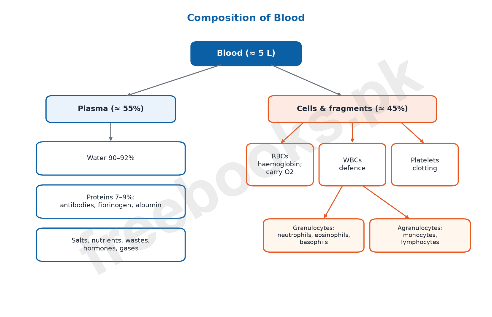
Blood is a fluid connective tissue. An adult has about 5 litres. About 55% of blood is plasma (the liquid part) and about 45% is cells and cell fragments.
Plasma is about 90–92% water, 7–9% proteins and about 1% other substances. Its proteins include antibodies (defence against germs), fibrinogen (needed for clotting) and albumin (maintains the osmotic pressure of blood). Plasma without fibrinogen is called serum.
Plasma also carries salts (mainly sodium, chloride and bicarbonate ions, with small amounts of calcium, potassium and others), digested nutrients such as glucose, amino acids and lipids, wastes such as urea, hormones from endocrine glands, and small amounts of dissolved oxygen and carbon dioxide.
3.1 Red Blood Cells
Red blood cells (erythrocytes) are biconcave discs with no nucleus or organelles in mature form, which leaves more room for haemoglobin. Haemoglobin carries oxygen (bright red when oxygenated, dark red when not) and a little carbon dioxide.
There are about 4 to 5.5 million RBCs in each cubic millimetre of blood. Before and just after birth they are made in the liver and spleen; in adults they are made in the red bone marrow of bones such as the sternum, ribs and vertebrae. Each lives about 120 days and old cells are removed by the liver and spleen.
3.1 White Blood Cells and Platelets
White blood cells (leukocytes) are colourless, irregular, larger and far fewer than RBCs (about 7,000 per mm³). They are made in red bone marrow and some mature in lymph nodes, tonsils, thymus or spleen. They defend the body against disease.
Granulocytes have granules in their cytoplasm: neutrophils engulf bacteria and clear dead cells; eosinophils fight parasites and take part in allergies; basophils release histamine, which causes inflammation. Agranulocytes have clear cytoplasm: monocytes become macrophages that engulf germs and debris, and lymphocytes make antibodies.
Platelets (thrombocytes) are small fragments of large bone-marrow cells, without a nucleus, living 7 to 12 days, about 250,000 per mm³. When a vessel is damaged they gather at the site and start the clotting process: fibrinogen is changed into threads of fibrin that form a net, trapping red cells to make a clot that stops bleeding.
3.2 Structure of the Heart
The heart is a muscular pump lying in the chest between the two lungs, beneath the sternum. It is enclosed in a sac, the pericardium, whose fluid reduces friction as the heart beats.
A wall called the septum divides the heart into left and right sides. Each side has an upper thin-walled atrium and a lower thick-walled ventricle. The left ventricle has the thickest wall because it pumps blood to the whole body.
Valves let blood flow one way only. The tricuspid valve (three flaps) lies between the right atrium and right ventricle; the bicuspid valve (two flaps) between the left atrium and left ventricle. Semilunar valves at the bases of the pulmonary artery and aorta stop blood flowing back into the ventricles.
3.2 Double Circulation and Heartbeat
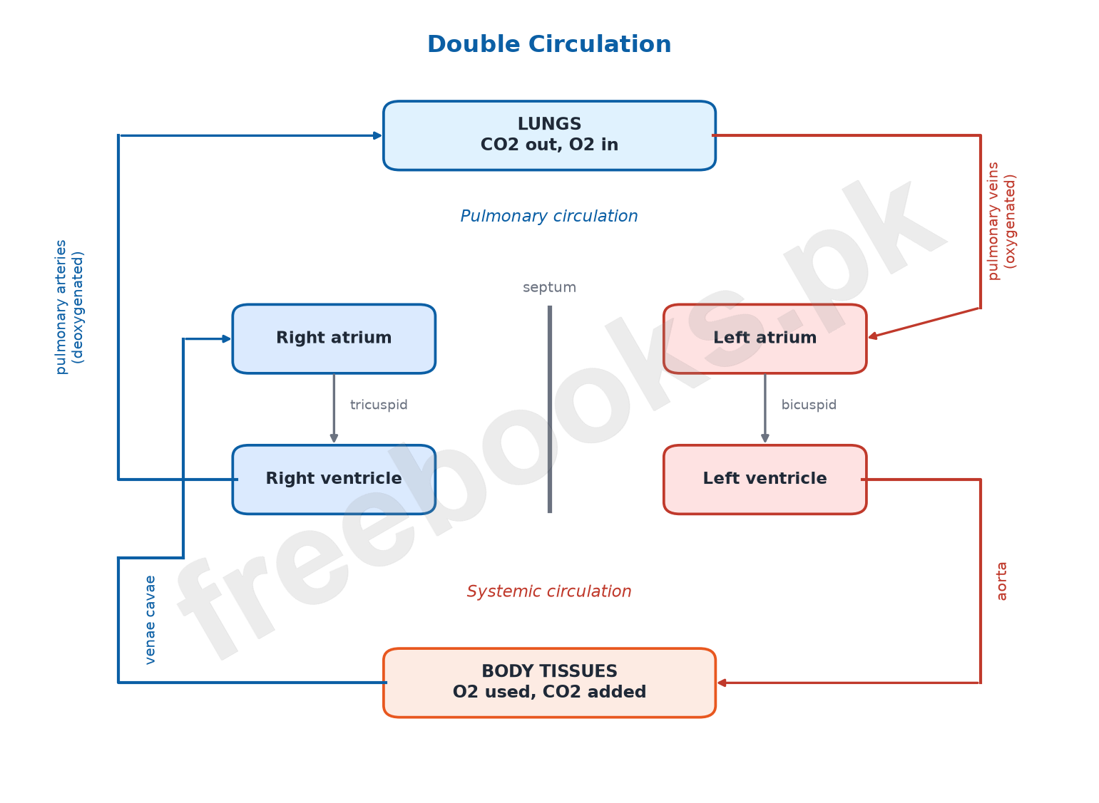
The superior and inferior venae cavae bring deoxygenated blood from the body into the right atrium → right ventricle → pulmonary arteries → lungs, where CO2 is lost and O2 is gained. Pulmonary veins return oxygenated blood to the left atrium → left ventricle → aorta → body.
Heart to lungs and back is pulmonary circulation; heart to body and back is systemic circulation. Because blood passes through the heart twice in one complete circuit, the heart works as a double pump. High pressure in the systemic circuit pushes blood to all organs; lower pressure in the lungs lets blood flow slowly, giving time for gas exchange.
One heartbeat is one contraction (systole) and relaxation (diastole) of the heart chambers; the average rate is about 70 beats per minute. During ventricular systole the ventricles pump blood into the arteries; in diastole the chambers relax and refill. The first sound ‘lubb’ comes from the closing of the tricuspid and bicuspid valves as the ventricles contract; the second sound ‘dubb’ comes from the closing of the semilunar valves.
3.3 Blood Vessels
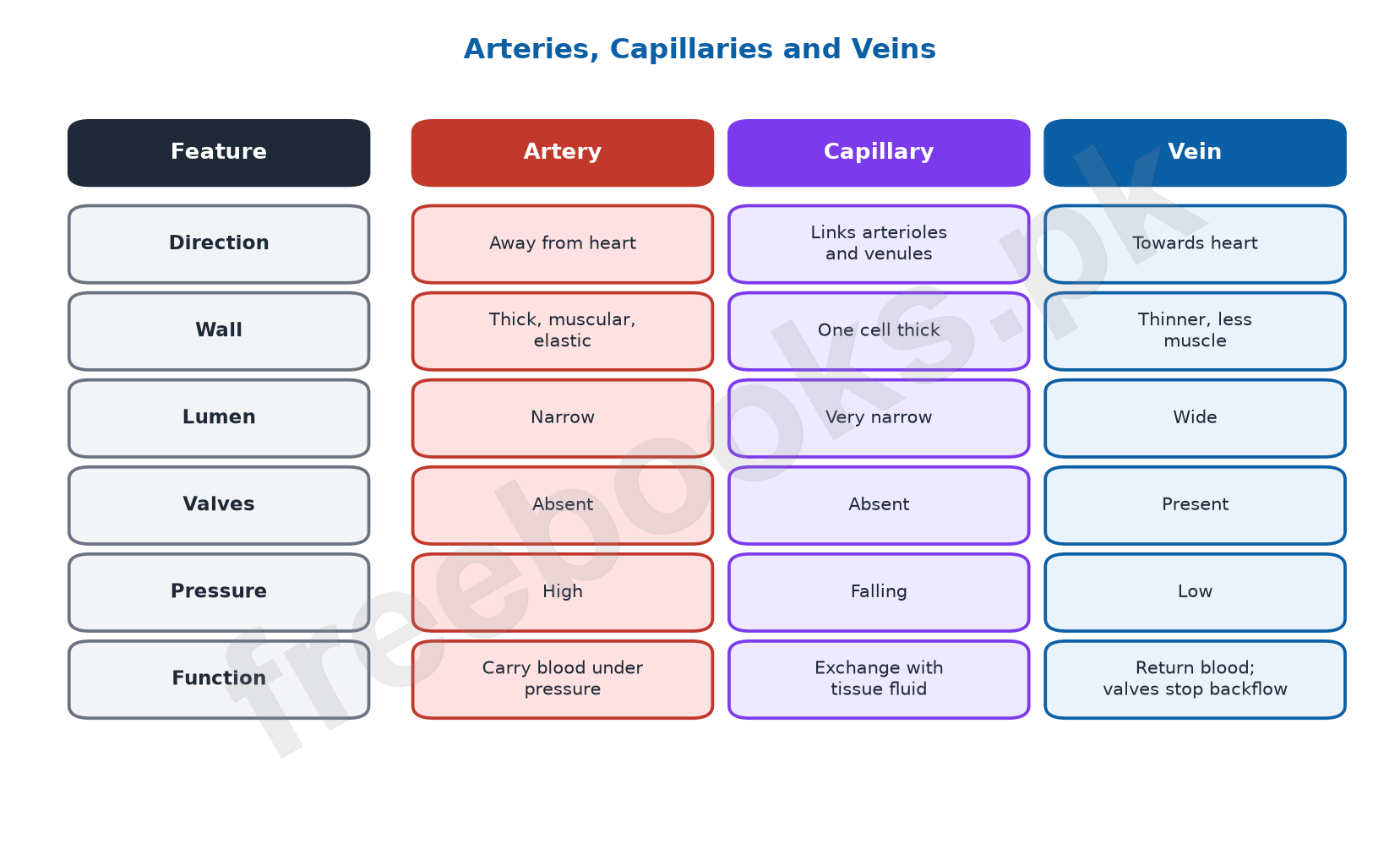
Arteries carry blood away from the heart (all carry oxygenated blood except the pulmonary arteries). Their thick, strong, elastic walls have three layers: inner endothelium, a middle layer of smooth muscle and elastic tissue, and an outer layer of connective tissue. They branch into arterioles and then capillaries.
Capillaries are the smallest vessels, with walls only one cell (endothelium) thick, so water, nutrients and oxygen pass out to the tissue fluid and carbon dioxide and wastes pass in. Capillaries join to form venules, which join to form veins.
Veins carry blood towards the heart (all carry deoxygenated blood except the pulmonary veins). They have the same three layers, but the middle layer is thinner, the lumen is wider, and most veins have valves that prevent backflow.
3.4 Arterial and Venous Systems
Arteries: the pulmonary trunk from the right ventricle splits into two pulmonary arteries, one to each lung. The aorta from the left ventricle gives coronary arteries to the heart muscle and branches to the head and arms, then runs down as the dorsal aorta, giving the hepatic artery (liver), renal arteries (kidneys) and finally the vessels that supply the legs (femoral arteries).
Veins: two pulmonary veins from each lung return blood to the left atrium. The superior vena cava drains the head, shoulders and arms; the inferior vena cava drains the lower body, receiving the femoral veins (legs), renal veins (kidneys) and the hepatic vein (liver). The hepatic portal vein carries blood from the gut to the liver. Coronary veins return blood from the heart muscle to the right atrium.
3.5 Cardiovascular Disorders
Coronary heart disease (CHD) is narrowing or blockage of the coronary arteries, mainly by atherosclerosis: fatty plaques containing cholesterol build up inside the arteries. Causes and risk factors include high LDL cholesterol, high blood pressure, smoking, diabetes, obesity, inactivity and family history. It may cause no symptoms at first, later chest pain. Prevention: healthy diet, exercise, no smoking and control of blood pressure and cholesterol.
Myocardial infarction (heart attack) is death of part of the heart muscle when its blood supply is blocked for a long time, usually by a clot forming on a plaque in a coronary artery. Symptoms: chest pain or pressure, breathlessness, sweating, pain spreading to the arm, neck or jaw; some attacks are ‘silent’. Treatment includes clot-dissolving medicines, angioplasty and stents. Complications: heart failure, arrhythmia and cardiac arrest.
Angina pectoris is chest pain or pressure caused by reduced blood flow to the heart muscle; it is a warning symptom of CHD. It is treated with lifestyle changes, medicines, angioplasty or stents, and if untreated can lead to a heart attack.
Smoking harms the heart by damaging artery linings and speeding up atherosclerosis, making platelets clump and clots form, narrowing vessels and raising blood pressure, increasing heart rate and causing arrhythmias, and letting carbon monoxide reduce the oxygen carried by haemoglobin.
Important Definitions
Plasma
The liquid part of blood, mostly water with dissolved substances.
Serum
Plasma from which fibrinogen has been removed.
Haemoglobin
The iron-containing red pigment in RBCs that carries oxygen.
Erythrocytes
Red blood cells.
Leukocytes
White blood cells that defend the body.
Macrophage
A large cell formed from a monocyte that engulfs germs and debris.
Platelets
Cell fragments that start blood clotting.
Fibrin
Insoluble protein threads that form the net of a blood clot.
Pericardium
The membrane sac around the heart.
Septum
The wall dividing the left and right sides of the heart.
Tricuspid valve
The three-flapped valve between the right atrium and right ventricle.
Bicuspid valve
The two-flapped valve between the left atrium and left ventricle.
Semilunar valves
Valves at the bases of the aorta and pulmonary artery.
Pulmonary circulation
Flow of blood from the heart to the lungs and back.
Systemic circulation
Flow of blood from the heart to the body and back.
Systole
Contraction phase of the heart.
Diastole
Relaxation phase of the heart, when chambers refill.
Lumen
The hollow space inside a blood vessel.
Atherosclerosis
Build-up of fatty plaque inside arteries.
Myocardial infarction
Death of part of the heart muscle; a heart attack.
Angina pectoris
Chest pain due to reduced blood flow to the heart muscle.
Key Facts at a Glance
| Item | Value / fact |
|---|---|
| Blood volume in an adult | About 5 litres |
| Plasma : cells | About 55% : 45% |
| Water in plasma | About 90–92% |
| RBC count | 4–5.5 million per mm³ |
| RBC life span | About 120 days |
| WBC count | About 7,000 per mm³ |
| Platelet count / life span | About 250,000 per mm³ / 7–12 days |
| Average heart rate | About 70 beats per minute |
| Thickest heart chamber | Left ventricle |
| Artery carrying deoxygenated blood | Pulmonary artery |
| Vein carrying oxygenated blood | Pulmonary vein |
| World Heart Day | 29 September |
Diagrams
Composition of blood: A chart of blood components: plasma and its contents, and the types of blood cells with their roles.
Double circulation: A flow diagram of blood through the right side of the heart to the lungs and the left side to the body.
Arteries, capillaries and veins: A comparison of the structure and function of the three types of blood vessels.
Short Questions and Answers
Why is blood called a connective tissue?
It consists of cells suspended in a liquid matrix (plasma) and links all parts of the body by transporting materials.
What is the difference between plasma and serum?
Serum is plasma without fibrinogen, so it cannot clot.
Give the roles of three plasma proteins.
Antibodies fight germs, fibrinogen helps blood clot, and albumin maintains the osmotic pressure of blood.
How is the shape of an RBC useful?
The biconcave disc shape gives a large surface for oxygen to diffuse in and out quickly.
Why do mature RBCs have no nucleus?
Lacking a nucleus and organelles leaves more space for haemoglobin, so each cell carries more oxygen.
Distinguish granulocytes from agranulocytes.
Granulocytes (neutrophils, eosinophils, basophils) have granules in their cytoplasm; agranulocytes (monocytes, lymphocytes) have clear cytoplasm.
What is pus?
A white fluid at an infection site made of dead white blood cells, dead germs and tissue debris.
How do platelets help stop bleeding?
They gather at a wound and start a process in which fibrinogen forms fibrin threads that trap blood cells into a clot.
Why is the wall of the left ventricle thicker than that of the right ventricle?
The left ventricle pumps blood around the whole body at high pressure, while the right pumps only to the nearby lungs.
What is the role of heart valves?
They let blood flow in one direction only and prevent backflow into the atria or ventricles.
Why is the human heart called a double pump?
Its right side pumps blood to the lungs and its left side pumps blood to the body, so blood passes through the heart twice per circuit.
What causes the ‘lubb’ and ‘dubb’ heart sounds?
‘Lubb’ is the closing of the tricuspid and bicuspid valves; ‘dubb’ is the closing of the semilunar valves.
Name an artery carrying deoxygenated blood and a vein carrying oxygenated blood.
The pulmonary artery carries deoxygenated blood; the pulmonary vein carries oxygenated blood.
Why do veins have valves but arteries usually do not?
Blood in veins is at low pressure and could flow backward; valves prevent this. High pressure in arteries keeps blood moving forward.
What is the difference between angina and a heart attack?
Angina is chest pain from reduced blood flow without lasting damage; in a heart attack the blood supply is blocked long enough for heart muscle to die.
Long Questions and Answers
Describe the composition of blood.
What are the main parts of blood?
About 55% plasma and 45% cells and cell fragments; an adult has about 5 litres.
What does plasma contain?
90–92% water, 7–9% proteins (antibodies, fibrinogen, albumin), and salts, nutrients, wastes, hormones and dissolved gases.
Describe red blood cells.
Biconcave cells without a nucleus, full of haemoglobin, 4–5.5 million per mm³, made in red bone marrow and living about 120 days.
Describe white blood cells and platelets.
WBCs (granulocytes and agranulocytes) defend the body; platelets are cell fragments that start clotting.
Describe the types of white blood cells and their functions.
What are neutrophils?
Granulocytes that engulf bacteria and clear dead cells at infection sites.
What do eosinophils and basophils do?
Eosinophils fight parasites and take part in allergies; basophils release histamine, causing inflammation.
What are monocytes?
Agranulocytes that become macrophages, which engulf germs and dead cells.
What do lymphocytes do?
They make antibodies against specific pathogens.
Describe the structure of the human heart.
Where is the heart and what covers it?
In the chest between the lungs, beneath the sternum, enclosed by the pericardium with fluid to reduce friction.
What are its chambers?
Two thin-walled atria above and two thick-walled ventricles below, separated into left and right sides by the septum.
Which valves are found between the chambers?
The tricuspid valve on the right and the bicuspid valve on the left, between each atrium and ventricle.
What are semilunar valves?
Valves at the openings of the pulmonary artery and aorta that stop blood returning to the ventricles.
Trace the path of blood through the heart, lungs and body.
How does blood enter the right side?
The superior and inferior venae cavae bring deoxygenated blood to the right atrium, which passes it to the right ventricle.
What is pulmonary circulation?
The right ventricle pumps blood through the pulmonary arteries to the lungs; oxygenated blood returns via pulmonary veins to the left atrium.
What is systemic circulation?
The left ventricle pumps oxygenated blood into the aorta and its branches to all body tissues; it returns via the venae cavae.
Why is this called double circulation?
Blood passes through the heart twice in one complete circuit; high systemic pressure reaches all organs while slower lung flow allows gas exchange.
Compare arteries, veins and capillaries.
Describe arteries.
Carry blood away from the heart; thick, elastic, muscular walls; narrow lumen; no valves (except at the heart).
Describe veins.
Carry blood to the heart; thinner middle layer; wide lumen; valves prevent backflow.
Describe capillaries.
Smallest vessels with walls one cell thick, allowing exchange between blood and tissue fluid.
Which vessels are exceptions in the oxygen they carry?
Pulmonary arteries carry deoxygenated blood and pulmonary veins carry oxygenated blood.
Describe coronary heart disease, heart attack and angina.
What is coronary heart disease?
Narrowing or blockage of the coronary arteries, mostly by atherosclerosis, linked to high cholesterol, high blood pressure, smoking, diabetes and inactivity.
What is myocardial infarction?
Death of part of the heart muscle when a coronary artery is blocked, often by a clot on a plaque; it causes chest pain, sweating and breathlessness.
How is a heart attack treated?
With clot-dissolving medicines, angioplasty to open the artery and stents to keep it open.
What is angina and how does smoking add to heart disease?
Angina is chest pain from reduced blood flow to the heart; smoking damages artery linings, promotes clots, raises blood pressure and heart rate, and lowers oxygen supply.
Multiple Choice Questions (MCQs)
The percentage of plasma in blood is about: (A) 45% (B) 55% (C) 75% (D) 90%
Correct answer: (B) 55%. About 55% plasma.
The plasma protein needed for clotting is: (A) Albumin (B) Fibrinogen (C) Antibody (D) Haemoglobin
Correct answer: (B) Fibrinogen. Fibrinogen forms fibrin.
Mature red blood cells lack a: (A) Cell membrane (B) Haemoglobin (C) Nucleus (D) Cytoplasm
Correct answer: (C) Nucleus. No nucleus, more room for haemoglobin.
The average life span of an RBC is about: (A) 7–12 days (B) 30 days (C) 120 days (D) 1 year
Correct answer: (C) 120 days. About 120 days.
Cells that release histamine are: (A) Neutrophils (B) Basophils (C) Monocytes (D) Lymphocytes
Correct answer: (B) Basophils. Basophils.
Antibodies are made by: (A) Lymphocytes (B) Platelets (C) RBCs (D) Eosinophils
Correct answer: (A) Lymphocytes. Lymphocytes make antibodies.
Platelets are best described as: (A) Complete cells (B) Cell fragments (C) Proteins (D) Antibodies
Correct answer: (B) Cell fragments. Fragments of large marrow cells.
The valve between the left atrium and left ventricle is the: (A) Tricuspid (B) Bicuspid (C) Semilunar (D) Pulmonary
Correct answer: (B) Bicuspid. Bicuspid has two flaps.
The chamber with the thickest wall is the: (A) Right atrium (B) Left atrium (C) Right ventricle (D) Left ventricle
Correct answer: (D) Left ventricle. It pumps to the whole body.
Blood from the lungs enters the heart at the: (A) Right atrium (B) Left atrium (C) Right ventricle (D) Left ventricle
Correct answer: (B) Left atrium. Via pulmonary veins.
The ‘dubb’ sound is caused by closing of the: (A) Tricuspid valve (B) Bicuspid valve (C) Semilunar valves (D) Septum
Correct answer: (C) Semilunar valves. Semilunar valves close.
Vessels with walls one cell thick are: (A) Arteries (B) Veins (C) Capillaries (D) Venae cavae
Correct answer: (C) Capillaries. Capillaries.
Most veins contain: (A) Thick muscle (B) Valves (C) Cartilage (D) Haemoglobin
Correct answer: (B) Valves. Valves stop backflow.
Blood from the gut is carried to the liver by the: (A) Hepatic artery (B) Hepatic vein (C) Hepatic portal vein (D) Renal vein
Correct answer: (C) Hepatic portal vein. Hepatic portal vein.
Death of heart muscle due to blocked blood supply is: (A) Angina (B) Atherosclerosis (C) Myocardial infarction (D) Arrhythmia
Correct answer: (C) Myocardial infarction. Heart attack.
Quick Revision Summary
- Blood ≈ 5 L: plasma 55% (water 90–92%, proteins, salts, nutrients, wastes, hormones, gases) + cells 45%.
- RBCs: biconcave, no nucleus, haemoglobin, 4–5.5 million/mm³, 120 days. WBCs: ~7,000/mm³; neutrophils, eosinophils, basophils, monocytes, lymphocytes. Platelets: 250,000/mm³, clotting.
- Heart: pericardium, septum, 2 atria + 2 ventricles; tricuspid (right), bicuspid (left), semilunar valves.
- Path: venae cavae → RA → RV → pulmonary arteries → lungs → pulmonary veins → LA → LV → aorta → body.
- Heartbeat ~70/min; systole/diastole; lubb = AV valves close, dubb = semilunar valves close.
- Arteries (thick, elastic, away from heart), capillaries (one cell thick, exchange), veins (thin, wide lumen, valves, to heart).
- CHD (atherosclerosis), myocardial infarction (heart attack), angina (chest pain). Smoking worsens all of them.
Exam Tips
- Practise tracing the path of blood through the heart in order; it is a frequent long question.
- Learn the exceptions: pulmonary artery (deoxygenated) and pulmonary vein (oxygenated).
- Draw and label the heart with chambers, valves and the four main vessels.
- Make a three-column table comparing arteries, veins and capillaries.
- Remember the numbers: 5 L blood, 55/45, 120 days, 7,000 WBCs, 250,000 platelets, 70 beats per minute.
Note on Facts
The textbook calls the sternum, ribs and vertebrae ‘short bones’. The sternum and ribs are flat bones and the vertebrae are irregular bones; the point to learn is that adult RBCs are made in the red marrow of these bones.
The textbook describes diastole as the relaxation of the atria. In diastole the ventricles relax as well and the whole heart refills with blood; contraction of the ventricles is systole.
The textbook says platelets convert fibrinogen into fibrin and that pus forms from dead macrophages. Platelets start a chain of reactions in which the enzyme thrombin converts fibrinogen to fibrin, and pus is made mostly of dead neutrophils along with germs and debris.