Chapter 1 follows food on its journey through the human body. Food contains large molecules (polymers) such as starch, proteins and fats that cannot pass through cell membranes. The digestive system breaks them into small soluble units (monomers) such as glucose, amino acids, fatty acids and glycerol, which are absorbed into blood and lymph and used by the cells.
These notes explain the five steps of nutrition, each region of the alimentary canal from the oral cavity to the anus, swallowing and peristalsis, the enzymes acting in each region, the structure of a villus, the many roles of the liver, and three common disorders of the gut: diarrhoea, constipation and peptic ulcer.
Learning Objectives
- Describe ingestion, digestion, absorption, assimilation and egestion.
- Describe the main regions of the alimentary canal and the accessory organs.
- Explain swallowing and peristalsis.
- State the enzymes of each region with their substrates and products.
- Describe the structure of a villus and the roles of capillaries and lacteals.
- State the roles of the liver and the causes, symptoms, treatment and prevention of diarrhoea, constipation and ulcers.
Key Concepts
1.1 Nutrition and Its Steps
Nutrition is the way an organism obtains and uses nutrients for energy, growth and repair. Plants show autotrophic nutrition: they make food from inorganic substances using light or chemical energy. Humans and other animals show heterotrophic nutrition: they obtain ready-made food from other organisms.
Nutrition in humans has five steps. Ingestion is taking food into the mouth. Digestion is breaking food down into small, soluble molecules. Absorption is the passage of these molecules into blood or lymph. Assimilation is the use of absorbed nutrients by body cells. Egestion is the removal of undigested waste through the anus.
Digestion is needed because carbohydrates, proteins and fats are large polymers that cannot cross cell membranes. They must be broken into monomers such as simple sugars, amino acids, fatty acids and glycerol.
1.2 Oral Cavity, Pharynx and Oesophagus
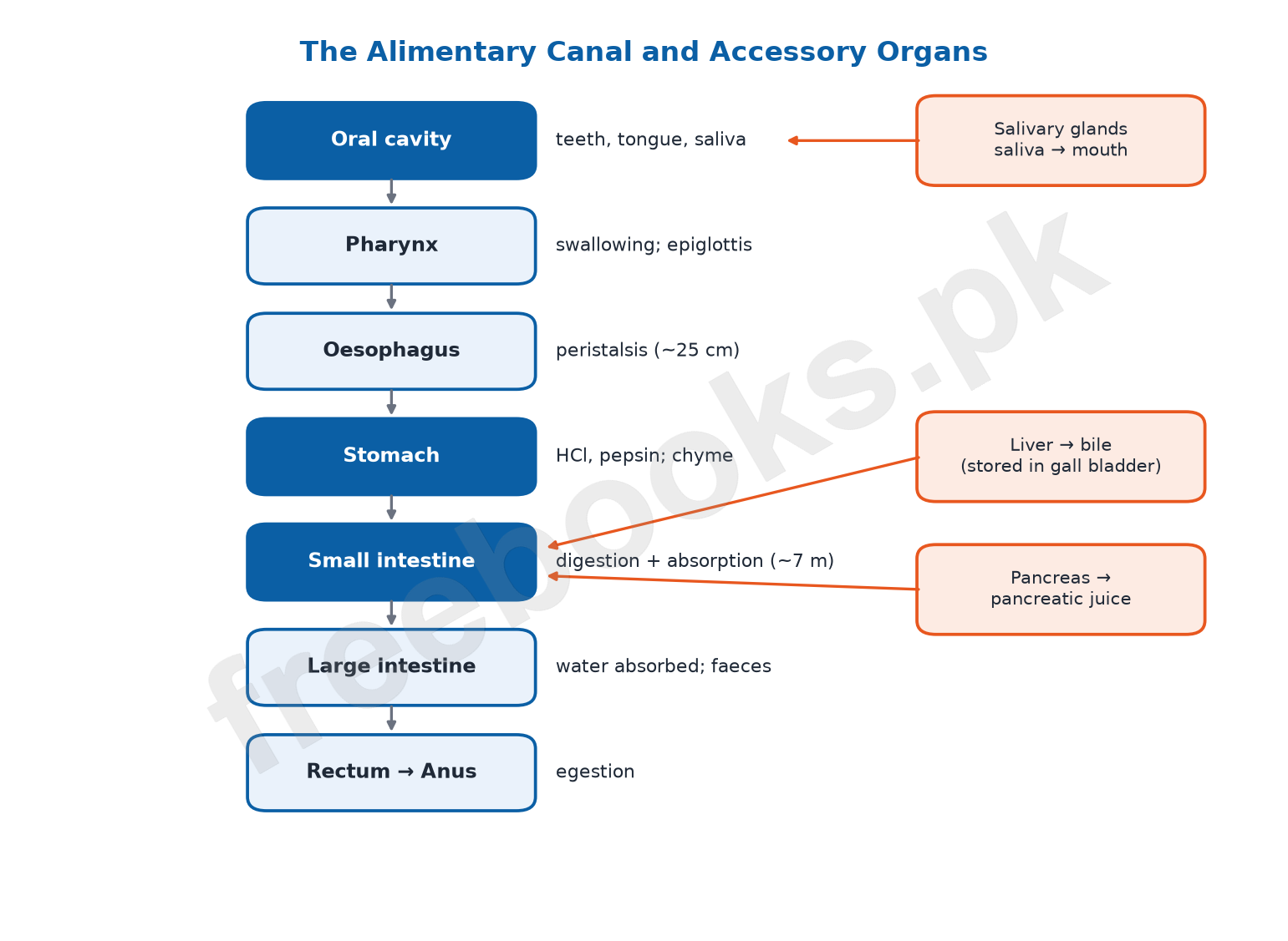
The alimentary canal is a long tube from mouth to anus. Accessory organs (salivary glands, liver and pancreas) pour their secretions into it.
In the oral cavity the teeth cut and grind food (mechanical digestion) while the tongue keeps it between the teeth; taste buds on the tongue detect taste. Three pairs of salivary glands secrete saliva, which contains water and mucus to moisten food and the enzyme salivary amylase, which begins the chemical digestion of starch into maltose.
Swallowing: the chewed food, now a soft ball called a bolus, is pushed by the tongue to the back of the mouth into the pharynx. The soft palate rises to close the nasal passage, and the larynx moves up against the epiglottis so the glottis is closed and food does not enter the trachea.
The oesophagus, a tube about 25 cm long, carries food to the stomach by peristalsis: waves of contraction and relaxation of the muscles in its wall that squeeze food along. No new digestive juice is added in the oesophagus.
1.2 Stomach
The stomach is a J-shaped muscular bag in the upper left abdomen below the diaphragm. Food enters at the cardiac end through the cardiac (lower oesophageal) sphincter, which stops food flowing back, and leaves at the pyloric end through the pyloric sphincter.
The thick muscular wall churns food and mixes it with gastric juice from gastric glands. Gastric juice contains hydrochloric acid, which kills many bacteria and converts inactive pepsinogen into active pepsin; pepsin breaks proteins into shorter chains called peptides. Mucus lines the wall and protects it from the acid and pepsin.
Food stays in the stomach for about three to four hours and becomes a thick soup-like liquid called chyme. The pyloric sphincter releases chyme into the small intestine a little at a time (about 5 to 15 mL at a time).
1.2 Small Intestine and Digestion
The small intestine is a coiled tube about 7 m long with three parts: duodenum (about 25 cm), jejunum (about 2.5 m) and ileum (about 4 m).
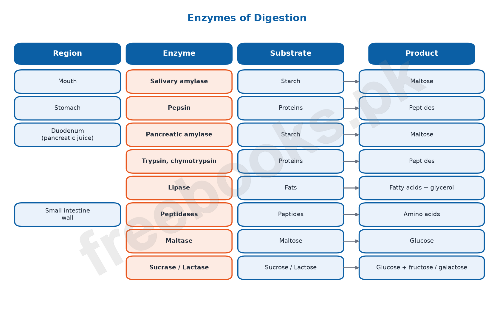
In the duodenum, bile from the liver arrives through the common bile duct. Bile has no enzymes, but its bile salts break large fat globules into tiny droplets (emulsification), giving lipase a larger surface to act on. Pancreatic juice arrives through the pancreatic duct; it contains pancreatic amylase (starch → maltose), trypsin and chymotrypsin (proteins → peptides) and lipase (fats → fatty acids and glycerol), as well as sodium bicarbonate, which neutralises the acid chyme.
Glands in the intestinal wall complete digestion: peptidases and dipeptidases change peptides into amino acids, maltase changes maltose into glucose, sucrase changes sucrose into glucose and fructose, lactase changes lactose into glucose and galactose, and nucleases break nucleic acids into nucleotides.
1.2 Absorption: Villi
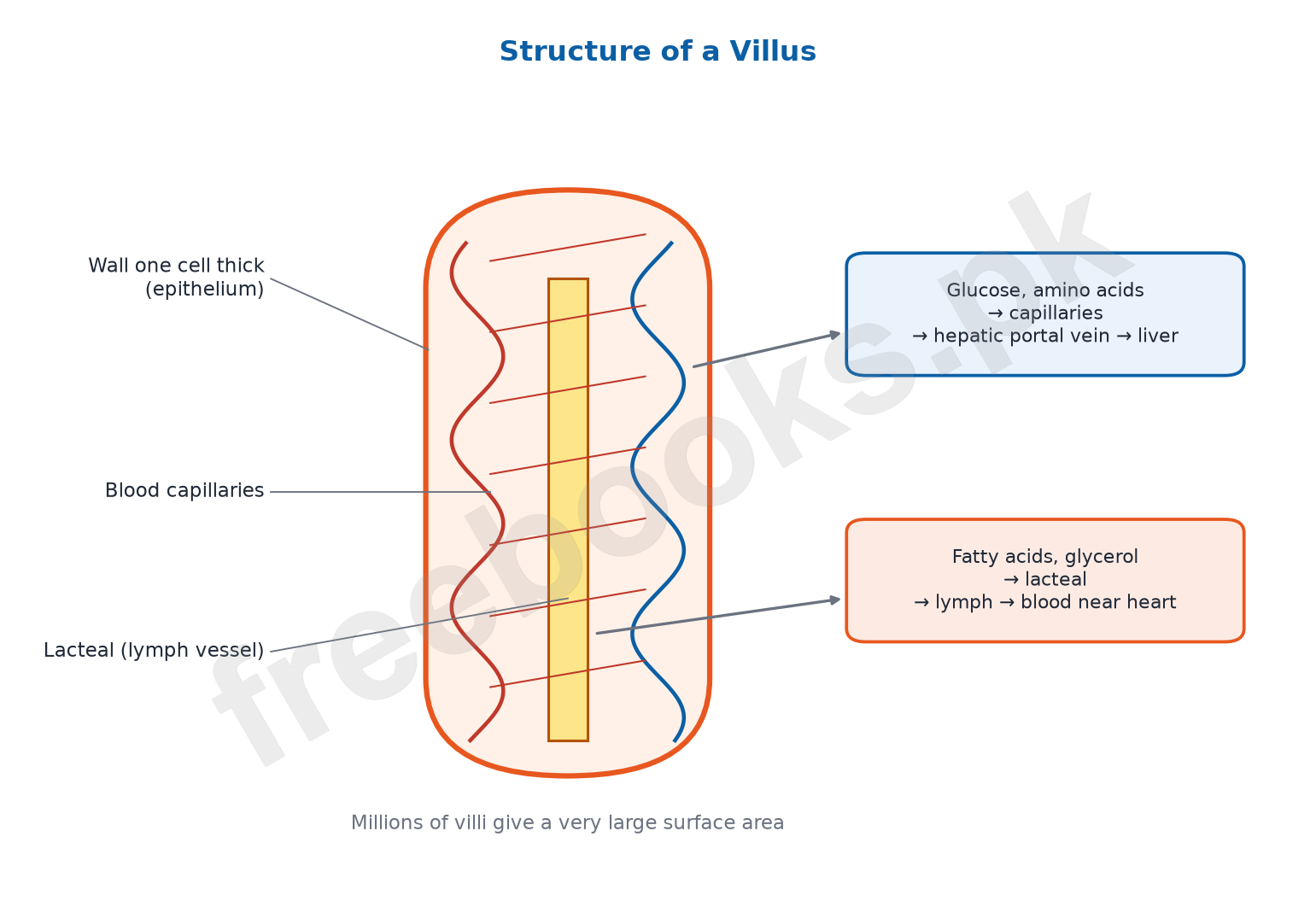
The inner wall of the small intestine is folded and covered with millions of finger-like villi, which give a very large surface area for absorption. The wall of each villus is only one cell thick, so the distance for absorption is short. Food molecules are absorbed by diffusion and active transport.
Each villus contains a network of blood capillaries and a central lymph vessel called a lacteal. Glucose and amino acids enter the capillaries, which join to form the hepatic portal vein carrying them to the liver; from there the hepatic vein carries them towards the heart. Fatty acids and glycerol enter the lacteal and travel through the lymph vessels, which empty into the blood near the heart.
1.2 Large Intestine
Undigested material passes into the large intestine, which has three parts: caecum, colon and rectum. A small finger-like appendix hangs from the caecum; if it becomes infected (appendicitis) and is not removed, it may burst and spread infection.
The large intestine absorbs most of the remaining water, so the waste becomes semi-solid faeces, containing undigested fibre, bacteria, dead cells from the gut lining, bile pigments and water. Faeces are stored in the rectum and expelled through the anus (egestion).
1.2 Role of the Liver
The liver is the largest gland, weighing about 1.5 kg in an adult, lying on the right side below the diaphragm. It makes bile, which is stored in the gall bladder and released into the duodenum. Bile pigments come from the breakdown of old red blood cells and leave the body in the faeces.
Other functions: it stores glucose as glycogen and releases glucose when needed; stores fat-soluble vitamins; breaks down excess amino acids and turns the toxic ammonia produced into less toxic urea; breaks down toxins such as alcohol; breaks down old red blood cells; makes vitamin A from carotene; and produces heat by its high rate of metabolism.
1.3 Disorders of the Digestive System
Diarrhoea is the passing of frequent, loose, watery stools. Symptoms include abdominal cramps, bloating and signs of dehydration (thirst, dry mouth, dark urine). Causes include bacterial, viral or parasitic infection, food allergy, stress and some medicines. Treatment is plenty of fluids and oral rehydration solution (ORS), soft food, and medicines or antibiotics in severe cases. Prevention: wash hands before eating and after using the toilet and avoid contaminated food and water.
Constipation is infrequent (fewer than three times a week) or difficult passing of hard, dry faeces, with discomfort or bloating. Causes include a low-fibre diet, too little water, lack of exercise, some medicines (such as painkillers and iron tablets) and some diseases such as diabetes. Treatment and prevention: eat more fibre (fruits, vegetables, whole grains), drink plenty of water, exercise regularly and take medicine only on a doctor’s advice.
A peptic ulcer is a sore in the lining of the stomach (gastric ulcer), duodenum (duodenal ulcer) or oesophagus. Symptoms include burning stomach pain, loss of appetite, weight loss and, in severe cases, vomiting blood. The main causes are infection by the bacterium Helicobacter pylori and long-term use of painkillers called NSAIDs; alcohol and smoking add to the risk and stress can worsen symptoms. Treatment uses antibiotics against H. pylori, antacids and acid-reducing medicines (proton pump inhibitors).
Important Definitions
Nutrition
The process by which an organism obtains and uses nutrients.
Heterotrophic nutrition
Obtaining ready-made food by consuming other organisms or organic matter.
Ingestion
Taking food and drink into the mouth.
Digestion
Breakdown of large food molecules into small soluble molecules.
Absorption
Passage of digested food molecules into the blood or lymph.
Assimilation
Use of absorbed nutrients by the body’s cells.
Egestion
Removal of undigested food from the body as faeces.
Bolus
A soft ball of chewed food ready to be swallowed.
Epiglottis
A flap of elastic cartilage that keeps swallowed food out of the trachea.
Peristalsis
Waves of muscular contraction that push food along the alimentary canal.
Sphincter
A ring of muscle that controls the opening of a tube.
Chyme
The soup-like mixture of partly digested food leaving the stomach.
Emulsification
Breaking of large fat globules into tiny droplets by bile salts.
Villi
Finger-like projections of the small intestine wall that absorb food.
Lacteal
The lymph vessel inside a villus that absorbs fatty acids and glycerol.
Hepatic portal vein
The vein that carries absorbed sugars and amino acids from the intestine to the liver.
Diarrhoea
Frequent passing of loose, watery stools.
Constipation
Infrequent or difficult passing of hard, dry faeces.
Peptic ulcer
A sore in the lining of the stomach, duodenum or oesophagus.
Key Facts at a Glance
| Region / organ | Key point |
|---|---|
| Salivary glands | 3 pairs; saliva with salivary amylase (starch → maltose) |
| Oesophagus | About 25 cm; moves food by peristalsis |
| Stomach | HCl + pepsinogen → pepsin (proteins → peptides); food stays 3–4 h |
| Small intestine length | About 7 m: duodenum 25 cm, jejunum 2.5 m, ileum 4 m |
| Bile | No enzymes; bile salts emulsify fats |
| Pancreatic juice | Amylase, trypsin, chymotrypsin, lipase + sodium bicarbonate |
| Intestinal enzymes | Maltase, sucrase, lactase, peptidases, nucleases |
| Sugars & amino acids absorbed into | Blood capillaries → hepatic portal vein → liver |
| Fatty acids & glycerol absorbed into | Lacteals (lymph) |
| Large intestine | Caecum, colon, rectum; absorbs water |
| Liver mass | About 1.5 kg |
| Main cause of peptic ulcer | Helicobacter pylori infection; long-term NSAIDs |
Diagrams
The alimentary canal: A flow chart of the alimentary canal from oral cavity to anus with the accessory organs and their secretions.
Enzymes of digestion: A chart of the main digestive enzymes in each region with their substrates and products.
Structure of a villus: A simplified villus showing its one-cell-thick wall, blood capillaries, lacteal and where each nutrient goes.
Short Questions and Answers
Why must food be digested before it can be used?
Starch, proteins and fats are large insoluble molecules that cannot cross cell membranes; digestion turns them into small soluble molecules that can be absorbed.
What is the difference between absorption and assimilation?
Absorption is the movement of digested food into the blood or lymph; assimilation is the use of these absorbed nutrients by the body cells.
What are the roles of saliva?
Its water and mucus moisten and lubricate food so it can be swallowed, and salivary amylase begins digesting starch into maltose.
How is food prevented from entering the windpipe?
During swallowing the larynx rises against the epiglottis, closing the glottis, so the bolus passes over it into the oesophagus.
Why can food reach the stomach even when a person is lying down?
Food is moved by peristalsis, muscular waves in the oesophagus wall, not by gravity.
Give two functions of hydrochloric acid in the stomach.
It kills many bacteria present in food and converts inactive pepsinogen into active pepsin.
Why does the stomach not digest itself?
A layer of mucus covers its inner wall and protects it from acid and pepsin; pepsin is also secreted in an inactive form.
What is the role of the pyloric sphincter?
It controls the release of chyme from the stomach into the duodenum, a small amount at a time.
How does bile help digestion although it has no enzymes?
Bile salts emulsify fats into tiny droplets, increasing the surface area on which lipase can act.
Why is sodium bicarbonate present in pancreatic juice?
It neutralises the acidic chyme from the stomach, giving the alkaline conditions intestinal enzymes need.
Name the enzymes that act on maltose, sucrose and lactose.
Maltase (maltose → glucose), sucrase (sucrose → glucose + fructose) and lactase (lactose → glucose + galactose).
How are villi adapted for absorption?
They give a huge surface area, have walls one cell thick, and contain many blood capillaries and a lacteal to carry absorbed food away.
Why can a person with liver disease have trouble digesting fats?
The liver makes bile; without enough bile, fats are not emulsified and lipase works much more slowly.
Why is ORS given in diarrhoea?
It replaces the water and salts lost in watery stools and prevents dehydration, which is dangerous especially for children.
How does a high-fibre diet prevent constipation?
Fibre adds bulk and holds water in the faeces, keeping them soft and helping peristalsis move them along.
Long Questions and Answers
Describe digestion in the oral cavity, pharynx and oesophagus.
What happens to food in the oral cavity?
Teeth cut and grind food while the tongue moves it; saliva from three pairs of salivary glands moistens it and salivary amylase begins breaking starch into maltose.
How is food swallowed?
The tongue pushes the bolus into the pharynx; the soft palate closes the nasal passage and the epiglottis covers the glottis so food goes into the oesophagus.
What is peristalsis?
Waves of contraction and relaxation of the circular and longitudinal muscles in the oesophagus wall that squeeze food down to the stomach.
Does chemical digestion continue in the oesophagus?
No new enzymes are added, but salivary amylase mixed with the bolus can continue to act on starch until stomach acid stops it.
Describe the structure and functions of the stomach.
Where is the stomach and what are its parts?
A J-shaped bag in the upper left abdomen below the diaphragm, with a cardiac end guarded by the cardiac sphincter and a pyloric end guarded by the pyloric sphincter.
How is food broken down mechanically?
Strong contractions of its thick muscular wall churn the food and mix it with gastric juice.
What does gastric juice do?
Its HCl kills bacteria and activates pepsinogen to pepsin, which digests proteins into peptides; mucus protects the stomach lining.
What leaves the stomach?
After 3–4 hours food becomes chyme, which the pyloric sphincter releases into the duodenum in small amounts.
Explain digestion in the small intestine and the role of the liver and pancreas.
What are the parts of the small intestine?
Duodenum (about 25 cm), jejunum (about 2.5 m) and ileum (about 4 m), making about 7 m in all.
What is the role of the liver in digestion?
It makes bile, stored in the gall bladder; bile salts emulsify fats into small droplets so lipase can digest them quickly.
What is the role of the pancreas?
It secretes pancreatic juice containing amylase, trypsin, chymotrypsin and lipase to digest starch, proteins and fats, plus sodium bicarbonate to neutralise chyme.
How is digestion completed?
Enzymes from the intestinal wall (peptidases, maltase, sucrase, lactase, nucleases) turn peptides and disaccharides into amino acids and simple sugars.
Describe the absorption of food in the small intestine.
How is the surface area increased?
The inner wall is folded and covered in millions of finger-like villi.
What is the structure of a villus?
A projection with a wall one cell thick, containing a network of blood capillaries and a central lacteal.
Where do sugars and amino acids go?
They enter the blood capillaries, which join the hepatic portal vein to the liver; from there the hepatic vein carries them towards the heart.
Where do fatty acids and glycerol go?
They enter the lacteal, travel through the lymph vessels and reach the blood near the heart.
State the roles of the liver in the body.
What is the liver’s role in digestion?
It makes bile, which emulsifies fats; bile pigments from broken-down red blood cells leave with the faeces.
How does it control food stores?
It stores glucose as glycogen and releases glucose when needed, and stores fat-soluble vitamins; it makes vitamin A from carotene.
How does it deal with wastes and toxins?
It breaks down excess amino acids, changes toxic ammonia into urea, breaks down alcohol and other toxins, and destroys old red blood cells.
How does it help keep the body warm?
Its high rate of metabolism releases a lot of heat, which increases in cold conditions.
Describe diarrhoea, constipation and peptic ulcer.
What are the causes and treatment of diarrhoea?
It is caused by infections, food allergy, stress or some medicines; treatment is fluids and ORS, soft food and medicine in severe cases.
What are the causes and prevention of constipation?
It results from low fibre, too little water, lack of exercise or some medicines; a high-fibre diet, plenty of water and exercise prevent it.
What causes a peptic ulcer?
Mainly Helicobacter pylori infection and long-term use of NSAID painkillers, with alcohol and smoking adding to the risk.
How is a peptic ulcer treated?
With antibiotics to remove H. pylori and antacids or proton pump inhibitors to reduce stomach acid.
Multiple Choice Questions (MCQs)
Using absorbed nutrients to build body cells is called: (A) Ingestion (B) Absorption (C) Assimilation (D) Egestion
Correct answer: (C) Assimilation. Assimilation is using absorbed food.
Salivary amylase converts starch into: (A) Glucose (B) Maltose (C) Sucrose (D) Peptides
Correct answer: (B) Maltose. Starch → maltose.
The flap that stops food entering the trachea is the: (A) Pharynx (B) Epiglottis (C) Palate (D) Sphincter
Correct answer: (B) Epiglottis. The epiglottis covers the glottis.
Food moves along the oesophagus by: (A) Gravity (B) Peristalsis (C) Diffusion (D) Emulsification
Correct answer: (B) Peristalsis. Muscular waves push food.
Pepsinogen is activated in the stomach by: (A) Bile (B) Mucus (C) HCl (D) Lipase
Correct answer: (C) HCl. HCl converts pepsinogen to pepsin.
The partly digested liquid leaving the stomach is: (A) Bolus (B) Chyme (C) Bile (D) Faeces
Correct answer: (B) Chyme. Chyme.
The longest part of the small intestine is the: (A) Duodenum (B) Jejunum (C) Ileum (D) Caecum
Correct answer: (C) Ileum. Ileum is about 4 m.
Bile helps digestion by: (A) Digesting proteins (B) Emulsifying fats (C) Digesting starch (D) Killing bacteria only
Correct answer: (B) Emulsifying fats. Bile salts emulsify fat.
Trypsin is secreted by the: (A) Stomach (B) Liver (C) Pancreas (D) Salivary glands
Correct answer: (C) Pancreas. It is in pancreatic juice.
Lactase breaks lactose into: (A) Two glucose (B) Glucose and fructose (C) Glucose and galactose (D) Amino acids
Correct answer: (C) Glucose and galactose. Lactose = glucose + galactose.
Fatty acids and glycerol are absorbed into the: (A) Hepatic portal vein (B) Lacteals (C) Capillaries only (D) Large intestine
Correct answer: (B) Lacteals. They enter the lymph in lacteals.
Most water from undigested food is absorbed in the: (A) Stomach (B) Large intestine (C) Oesophagus (D) Duodenum
Correct answer: (B) Large intestine. The colon absorbs water.
The liver converts toxic ammonia into: (A) Glycogen (B) Urea (C) Bile salts (D) Vitamin A
Correct answer: (B) Urea. Urea is less toxic.
A common bacterial cause of peptic ulcer is: (A) E. coli (B) Helicobacter pylori (C) Salmonella (D) Rhizopus
Correct answer: (B) Helicobacter pylori. H. pylori infects the stomach lining.
ORS is mainly used to treat dehydration caused by: (A) Constipation (B) Diarrhoea (C) Ulcer (D) Appendicitis
Correct answer: (B) Diarrhoea. It replaces water and salts.
Quick Revision Summary
- Steps: ingestion → digestion → absorption → assimilation → egestion.
- Mouth: teeth + tongue (mechanical); saliva with salivary amylase (starch → maltose). Swallowing: bolus → pharynx; epiglottis closes glottis.
- Oesophagus (~25 cm): peristalsis. Stomach: churning; HCl + pepsin (proteins → peptides); mucus protects; chyme; cardiac and pyloric sphincters.
- Small intestine (~7 m: duodenum, jejunum, ileum): bile emulsifies fats; pancreatic amylase, trypsin, chymotrypsin, lipase, NaHCO3; intestinal maltase, sucrase, lactase, peptidases.
- Villi: large surface, one-cell wall; capillaries (sugars, amino acids → hepatic portal vein → liver), lacteal (fatty acids, glycerol → lymph).
- Large intestine: caecum, colon, rectum; absorbs water; faeces egested.
- Liver: bile, glycogen store, vitamins, urea from ammonia, detoxification, breaks old RBCs, vitamin A, heat.
- Diarrhoea (ORS), constipation (fibre, water), peptic ulcer (H. pylori, NSAIDs; antibiotics, antacids).
Exam Tips
- Learn the enzyme table (region – enzyme – substrate – product); it gives many MCQs and short questions.
- Remember: bile has NO enzyme. Questions often test this.
- Know where each nutrient is absorbed: sugars and amino acids into blood, fatty acids and glycerol into lacteals.
- Draw a labelled villus and a labelled digestive system; both are common drawing questions.
- For disorders, answer under four headings: symptoms, causes, treatment, prevention.
Note on Facts
The textbook says heat produced by stomach churning helps melt lipids. Fats in food are kept fluid mainly by normal body temperature; the heat from churning is very small.
The textbook lists ‘avoiding spicy foods’ as prevention of ulcers. Spicy food does not cause peptic ulcers, though it can make the pain of an existing ulcer worse. The main causes are H. pylori infection and long-term NSAID use.
The textbook says mucus neutralises the stomach’s acid. More precisely, the mucus layer traps bicarbonate secreted by the lining, which neutralises acid right next to the wall while the stomach contents stay strongly acidic.